


A patient was referred to my office after receiving treatment from various providers with no results. The patient was training for the Olympics as a marathon runner and was unable to run or walk without severe medial knee pain.
After releasing the sartorius dysfunction, I had the patient test out his knee for pain and weakness. He was astounded that a simple fix of his hip flexor eliminated his knee pain in one visit. He finished his Olympic marathon training and then decided to go to chiropractic school himself. He is now practicing in California with great success.
Presentation
The sartorius muscle is a hip flexor and lower-leg external rotator. It originates along the medial border of the anterior surface of the anterior superior iliac spine and inserts into the pes anserine at the medial tibia. It can be strained, damaged and weakened through traumas such as stepping in a hole, slipping on ice, or tripping while walking. Patient complaints include medial knee pain, abnormal gait (aka, hip pointer), leg / foot drags or feels heavy, and/or inner-thigh discomfort. Onset of pain is typically acute, but may have a gradual progression.
Diagnosis
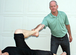 Diagnosing sartorius deficiency by pulling on the ankle to straighten the bent hip and knee.
With the patient laying supine, have them bend their knee to 90 degrees. Grasp their ankle and pull to straighten the bent hip and knee. Muscle test the sartorius bilaterally and note the deficiency of the involved sartorius.
Diagnosing sartorius deficiency by pulling on the ankle to straighten the bent hip and knee.
With the patient laying supine, have them bend their knee to 90 degrees. Grasp their ankle and pull to straighten the bent hip and knee. Muscle test the sartorius bilaterally and note the deficiency of the involved sartorius.
Treatment
There are several approaches to correcting the sartorius: instrument adjusting, pin and stretch techniques, and joint distraction / decompression can all be utilized to obtain results.
Adjusting instruments can target the insertion points. Pin and stretch protocols: Apply pressure into the insertions, trigger points and/or fibrous adhesions in the muscle belly, and then have the patient flex and extend their leg. Distraction / decompression is performed with the patient supine. Contact around the ankle and create traction through the entire leg, including the hip joint. Once traction is established, additional tractioning force (a gentle tug) can be added to unlock and distract the hip joint. Release of hip-joint tension can be felt by the doctor and patient as the femur head pulls slightly out of the acetabulum.
Posttreatment evaluation should note increased strength and range of motion function, and decreased pain.
Rehabilitation
Lunge stretches, performed slowly, can help reduce muscle tension and restore mobility. Strengthening exercises for the hip flexors can be incorporated to build endurance and stability. I prefer that my patients work the good side first and then train the injured side.
Dr. Todd Turnbull, has authored online courses and articles about concussions, sports performance, soft-tissue diagnosis, rehabilitation and disc herniations. He is a 1991 graduate of Life University, a board-certified chiropractic sports physician, and maintains a private practice in Portland, Ore. He can be contacted with questions or comments via his Web site: www.drtoddturnbull.com/DCJournal.