


Modern care focuses on the prevention of deconditioning and early patient reactivation, replacing the orthopedic approach, which focused on rest, medication and surgery. This biopsychosocial model is patient-centered, in that the focus of care is on reducing the patient's symptoms, distress and dysfunction.
Certain action steps are required to guide patients through the functional reactivation process. The first is to identify the patient's history of activity intolerances; the second is to assess the patient's "functional range" (FR); the third is to reactivate the patient through appropriate advice, manipulation and exercise; and the fourth is to reassess the patient's activity intolerances and FR through ongoing audit of outcomes.
Functional Reactivation Action Steps
- History of activity intolerances
- Assessment of the patient's "functional range"
- Functional reactivation
- Audit
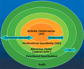
To carry out these action steps in patient care, we are faced with a series of clinical challenges. Each presents the chiropractor with a way to "operationalize" the goal of functional reactivation. The basic clinical questions are:
- What are the patient's activity intolerances?
- What are the patient's mechanical sensitivities?
- What is the relevant dysfunction or chain of dysfunction responsible for the patient's symptoms?
- Can you prescribe treatment for an acute patient that will expand his or her "functional range?"
- Can you isolate the patient's stabilization musculature?
- Can you prescribe functionally integrated training that re-educates appropriate movement patterns while simulating the patient's activities?
1. What Is the History of Activity Intolerances?
Clinical Challenge: To uncover (from the patient's history) what specific activity intolerances are present.
Record the patient's history of activity intolerance (AI). Use questionnaires such as the Oswestry or Neck Disability Index to quantify this. Use the patient's AI as an outcome and goal of care. For instance, if the patient has an aggravation of pain with prolonged sitting, reducing sitting sensitivity, as mutually agreed upon, is an excellent goal of care. Use the AI as a pretreatment audit on each follow-up visit, and re-evaluate quantifiable AI outcomes every 4-6 weeks.
2. Assessment of Functional Range
According to Dennis Morgan, PT, DC, a specialist in spinal stabilization exercises, "The functional range is the painless range which is appropriate for the task at hand." The FR has two components: One is the patient's mechanical sensitivity; the other is his or her abnormal motor control.
a. Mechanical Sensitivity (MS)
Clinical Challenge: To uncover (from the patient's examination) what specific movements and positions provoke or peripheralize symptoms.
Ask yourself if you can reproduce your patient's symptoms ("provocative testing") with any of the following tests:
- ROM
- Neural provocation (i.e. Butler's tests of adverse neural tension)
- Orthopedic
- Myofascial trigger points
- Provocative movements or positions (i.e., Kemps, Gaenslens)
Use the MS tests as an outcome and goal of care, and for pretreatment and posttreatment audit of patient progress. For instance, if active lumbar extension is the primary MS at the start of the visit, after manipulation or other therapy/exercise, retest the patient's sensitivity to back-bending to show the patient his or her improvement.
b. Functional Pathology or Abnormal Motor Control (AMC)
Clinical Challenge: To uncover (from the patient's examination) the key dysfunction or chain of dysfunction responsible for his or her symptoms or activity intolerance.
Abnormal motor control (AMC) is a typical signature of painful joint disorders. In the low-back-altered muscle, coactivation patterns between agonists and antagonists have been shown to be associated with changes in the back's response to lifting challenges, extremity motions or unexpected torso perturbations (Cholewicki, Hides, Hodges, Radebold). Specifically, delays in muscle activation and relaxation have been seen in back-pain patients. Similarly, in the cervical spine, the deep neck flexors' control of progressively increasing ranges of cervicocranial flexion is disturbed in whiplash or headache patients (Jull).
Joint blockage with pathokinesiology leads to AMC. In patient assessment, the clinician's goal is to find the source of the patient's pain. As chiropractors, we prefer to treat the cause of the pain, rather than just offering palliative care. For this reason, B.J. Palmer focused us on the upper-cervical region, and Logan did the same for the sacrum area. Neurologist Karel Lewit expanded this notion by teaching that various areas can be a "key link," such as the upper cervical, TMJ, pelvis or feet, and that "He who treats the site of pain is lost."
Various screening tests can be performed to detect AMC of the locomotor system. To efficiently prescribe reactivation training, the clinician needs to master the discreet set of individual tests listed here:
- Respiration
- Posture
- Arm elevation
- Wall angle
- Balance
- Single leg standing
- Vele's reflex stability test
- Core Stability
- Vleeming SLR
- Janda's hip extension and abduction
- Abdominal wall
- Abdominal hollowing
- Curl-up
- Trunk extensor endurance
- Side bridge endurance
- Cervico-cranial flexion
- Scapulo-thoracic
- Scapulo-humeral rhythm
- Push-up
- Squats/Lunges
- One- and two-leg squats
- Forward and angle lunges
3. Functional Reactivation
Clinical Challenge: To prescribe treatment for an acute patient that will expand his or her "functional range."
There are three steps to functional reactivation - advice, manipulation, and exercise.
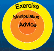
a. Advice
A patient with acute pain requires reassurance that he or she does not have a serious disease. As soon as "red flags" can be ruled out, the patient should be vigorously reassured that the pain is not due to anything serious. Patients also tend to assume they are injured or damaged; however, most back and neck pain is not due to an injury, but is likened more to an illness, such as a common cold. Therefore, patients should not be labeled as having a ruptured disc or degenerative arthritis, but be informed that their pain is due to a mechanical overload and will run a course similar to a cold (Bogduk; Indahl).
If patients have excessive anxiety or fear-avoidance behavior, labeling a condition will make them overly cautious with their backs. This is what is called a "yellow flag," in that it will slow recovery. Therefore, simple reactivation advice, such as encouragement to walk or perform light exercise, and that "hurt does not necessarily equal harm," is extremely important in minimizing the deconditioning that accompanies excessive fear-avoidance behavior (Vlaeyen).
Along with reassurance and reactivation advice, patients benefit from ergonomic advice regarding activity modifications, such as "Learn to roll out of bed instead of sitting up," or "Learn to hinge from the hips, rather than the waist, when rising from a chair or bending to the floor." According to Dr. Lewit, "The first treatment is to teach the patient to avoid what harms. ..." This can mean advising the patient not to do too little or too much!
b. The Role of Manipulation
As every chiropractor knows, if a joint is dysfunctional, adjusting it will reduce activity intolerances, mechanical sensitivity, and improve motor control. Naturally, the role of manipulation is to find those "key links" responsible for biomechanical overload in the entire kinetic chain. Such key dysfunctions are not limited to articular restrictions, but can include problems with the fascia, or even muscles (Lewit). Sometimes, an old scar is working as a saboteur, causing altered afferent input into the spinal cord. Whatever the "key link," manipulation of this peripheral source of abnormal neural input should be a primary option in patient care. In the report of findings, such manipulation should be viewed as a catalyst to reactivation, with manipulation viewed as a means, and reactivation as the end.
To empirically adjudicate that a "key link" has been found and remediated, always retest the patient's FR posttreatment. If manipulation is successful, find a self-treatment that reproduces the effects of the manipulation. For example, if low-back pain is secondary to a foot problem, manipulation of the foot will lead to improvement in the patient's mechanical sensitivity in his or her back. In this case, a home exercise, such as training of balance, or longitudinal and transverse arches of the foot, may achieve an effect similar to that of manipulation, and should therefore be prescribed for home treatment.
With exercise, it is important to bear in mind the object of care. The method of exercise should serve goals:
- Reduce activity intolerances
- Facilitate recovery from injury
- Train a person for a chosen task
- Prevent injury
- Enhance performance
As Dr. Lewit has noted, "remedial exercise is always time-consuming, and time should not be wasted. We should not attempt to teach patients ideal locomotor patterns, but only correct the fault that is causing the trouble."
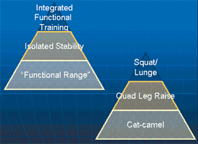
A stepwise approach to introducing exercise into patient care is to progress from exercises within the patient's FR to activation and isolation of the deep stabilizers, eventually including integration of the "core" in functional activities.
Exercise Progressions
- Functional range
- Isolated stability training
- Functionally integrated training (FIT)
1. Functional Range Exercises
Functional range exercises include gentle movements such as the cat-camel or BruŸger relief position, performed in the patient's most comfortable position or range. The goal is to improve pain-free ROM, so that training can be progressed.
2. Isolated Stability Training
Clinical Challenge: To isolate the patient's stabilization musculature.
Stability training should isolate the "deep" stabilizers responsible for maintaining integrity of the joints during loaded tasks. The ideal of such training is that it should maximally challenge the muscles while minimally stressing the joints (McGill). Examples include the quadruped leg reach for the multifidus, or cervicocranial flexion, such as nodding, for the deep neck flexors. The goal should be that MS decreases and AMC improves.
3. Functionally integrated training (FIT)
Clinical Challenge: Make training as functional as possible, so it will stabilize the patient in his or her home, sport and occupational activities.
Once stabilization musculature has been successfully isolated and the patient's FR is expanding, it is essential that exercises are progressed to include upright functional activities. These can include squats, lunges, and various triplaner pushing (punches) and pulling ("lawnmower") movements. The clinician should find the most functional activities within the patient's FR, and prescribe these. Physical therapist and authority on "humanomics," Gary Gray, terms this "attacking success." The goal is that the patient's FR expand, so that it encompasses the patient's activities of daily living, occupational demands and sports or recreational activities.
4. Audit
Treatment results should always be audited for continuous quality assurance. For chiropractic care to be a value-added approach to the health-care consumer, ongoing outcomes assessment of the patient's goals must be monitored regularly. One such audit question is, "Does manipulation or exercise of key functional pathologies improve the patient's functional range?" To find out, retest the patient's MS and AMC posttreatment; this will help adjudicate empirically the value of the treatment in the patient's eyes. It also will help motivate the patient, especially if the audit is performed after exercise. Another audit is to recheck activity intolerances at the start of each subsequent visit.
Conclusion
Can we identify our patients' relevant activity intolerances, mechanical sensitivities, and abnormal motor control? Can we provide treatments that expand our patients' "functional range?" Can we prescribe exercises that reeducate appropriate movement patterns, while simulating patients' activities? Do we regularly audit our patients' symptoms, activity intolerances (disability) and impairments (MS and AMC)?
References
- Bogduk N. What's in a name? The labeling of back pain. Medical Journal of Australia 2000;173:400-1.
- Cholewicki J, McGill SM. Mechanical stability of the in vivo lumbar spine: Implications for injury and chronic low back pain. Clin. Biomech 1996;11(1):1-15.
- Hides JA, Stokes MJ, Saide M, Jull GA, Cooper DH. Evidence of lumbar multifidus muscle wasting ipsilateral to symptoms in patients with acute/subacute low back pain. Spine 1994;19:165-172.
- Hides JA, Jull GA, Richardson CA. Long-term effects of specific stabilizing exercises for first-episode low back pain. Spine 2001;26:e243-e248.
- Hides JA, Richardson CA, Jull GA. Multifidus muscle recovery is not automatic after resolution of acute, first episode of low back pain. Spine 1996;21(23):2763-2769.
- Hodges PW, Richardson CA. Altered trunk muscle recruitment in people with low back pain with upper limb movement at different speeds. Arch Phys Med Rehabil 1999;80 (9):1005-12.
- Hodges PW, Richardson CA. Delayed postural contraction of transversus abdominis in low back pain associated with movement of the lower limb. J Spinal Disord 1998;11(1):46-56.
- Indahl A, Haldorsen EH, Holm S, Reikeras O, Hursin H. Five-year follow-up study of a controlled clinical trial using light mobilization and an informative approach to low back pain. Spine 1998;23:2625-2630.
- Lewit K. Manipulative Therapy in Rehabilitation of the Motor System, 3rd edition. London: Butterworths, 1999.
- McGill S. Stability: from biomechanical concept to chiropractic practice. J Can Chiro Assoc 1999;43:75-87.
- Radebold A, Cholewicki J, Panjabi MM, Patel TC. Muscle response pattern to sudden trunk loading in healthy individuals and in patients with chronic low back pain. Spine 2000;25:947-954.
- Radebold A, Cholewicki J, Polzhofer BA, Greene HS. Impaired postural control of the lumbar spine is associated with delayed muscle response times in patients with chronic idiopathic low back pain. Spine 2001;26:724-730.
- Vlaeyen JWS, Linton S. Fear-avoidance and its consequences in chronic musculoskeletal pain. A state of the art. Pain 2000;85:317-332.
Craig Liebenson, DC
Los Angeles, California
![]()
Click here for previous articles by Craig Liebenson, DC.