There are several normal anatomical differences between the adult and the pediatric cervical spine. This is a brief review of the main radiographic features that one should be aware of in order to avoid confusing normal differences with pathologic findings.
A child's spine is hypermobile compared to an adult's because of the relative ligamentous laxity, shallow and angled facet joints, underdeveloped spinous processes, and physiologic anterior wedging of vertebral bodies. Depending on the age of the child, there may be incomplete ossification of the odontoid process; add to this the relatively large head and weak neck muscles, and the pediatric spine is predisposed to increased instability compared to the adult spine.
The interpretation of pediatric cervical spine X-rays can be challenging, even for the most experienced, because of the wide range of normal anatomic variants and changes that occur with the maturation / ossification process. Therefore, familiarity with the normal anatomy will help avoid misinterpretations.
Embryonic Development
The first two cervical vertebrae are unique. C1 is formed by three primary ossification sites, the anterior arch and the two neural arches, which fuse to form the posterior arch. The anterior arch is ossified only in 20 percent of children at birth; with the other 80 percent it becomes visible as an ossification center by 1 year of age. The neural arches appear at the seventh fetal week. The anterior arch fuses with the neural arches by 7 years of age; nonfusion should not to be mistaken as a fracture. The neural arches fuse posteriorly by 3 years of age.
C2 is the most complex developmental and ossification process of all the vertebrae. There are four ossification centers at birth: one for each neural arch, one for the body and one for the odontoid process. The odontoid process forms in utero from two separate ossification centers that fuse in the midline by the seventh fetal month. A secondary ossification center appears at the apex of the odontoid process between 3-6 years of age. This fusion line can be seen until age 11 years and should not be confused with a fracture. The neural arches fuse posteriorly by 2-3 years of age and with the body of the odontoid process between 3-6 years of age.
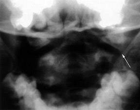
 Figure 1: The space between the lateral masses relative to the dens can be < 6 mm, but the lateral masses of C1 can still remain aligned with the lateral masses of C2 in the pediatric spine.
The vertebrae of C3 through C7 can be discussed as a group because they demonstrate the same developmental pattern. There are three primary ossification sites for each vertebral segment: the vertebral body, which develops from a single ossification center, and two neural arches. The neural arches fuse posteriorly by age 2-3 years and the body fuses with the neural arches between 3-6 years of age. In addition, there are secondary ossification centers at the tips of the transverse processes and spinous processes that may persist until early in the third decade of life, simulating a fracture. The secondary ossification centers at the superior and inferior aspects of the cervical vertebral bodies, the endplates, can remain unfused until early adulthood.
Normal Anatomic Variances
There are several normal anatomic differences that may be encountered on the pediatric cervical spine. One should be cognizant of these differences to avoid confusing them with pathologic conditions.
Atlantodens Interval (ADI): The ADI is defined as the distance between the anterior aspect of the dens and the posterior aspect of the anterior ring of the atlas. A normal ADI confirms that the transverse ligament is intact. This distance should be 5 mm or less in the pediatric cervical spine and not more than 3 mm in the adult.
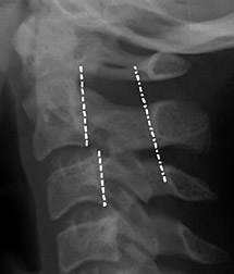
 Figure 2: Pseudo subluxation at the C2-3 level; note that the spinolaminar line remains contiguous.
Distance Between the Dens and Lateral Masses (Pseudo-Jefferson Fracture): The pseudo-spread of the atlas on the axis (pseudo-Jefferson fracture) is seen on the APOM view. Displacement up to 6 mm of the lateral masses relative to the dens is common up to the age of 4 years old, but can be seen up to 7 years of age. (Figure 1)
Pseudosubluxation at the C2-3 Level: In children, C2 can be displaced anteriorly in relation to C3 and to a lesser extent at the C3-4 level, resembling ligamentous injury. This finding is not uncommon in children less than 8 years of age, but may even be present in 20 percent of children under 16 years of age. It is caused by the horizontal nature of the facets joints in younger ages; as the spine matures the facet joints become more vertical.
True ligamentous injury can be determined by using the posterior cervical line, which is a line drawn from the anterior aspect of the spinous process of C1 to the anterior aspect of the spinous process of C3 (spinolaminar line). The anterior edges of the spinous processes of C1, C2 and C3 should line up within 1 mm of each other on both flexion and extension X-rays. If this line does not overlap the anterior aspect of the spinous process of the C2 by 2 mm or more, a true injury is present. (Figure 2)
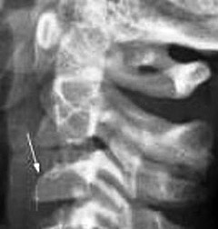
 Figure 3: Anterior wedging of the C3 vertebral body; a normal finding in the pediatric spine.
Absence of Lordosis: The absence of lordosis, although potentially pathologic in an adult, can be seen in children up to 16 years of age when the neck is in a neutral position. The normal posterior intraspinous distance is a good indicator of ligamentous integrity and should not be more than 1.5 times greater than the intraspinous distance one level either above or below the level in question. In children, the flexion maneuver can increase the distance between the tips of the C1 and C2 spinous processes. This widened C1-2 intervertebral distance is a normal finding and should not be misinterpreted as ligamentous injury. This finding is postulated to be secondary to the tight ligamentous attachment between the skull base and C1.
Secondary Ossification Centers: Knowing where the ossification centers appear is important in order not to confuse them with a fracture. In particular, the secondary centers of ossification of spinous processes and unfused ring apophyses of vertebral bodies can be confused with fractures. The normal physis is smooth, regular with a subchondral sclerotic line delineating the growth plate. Acute fractures can appear anywhere and are irregular, not sclerotic.
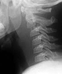
 Figure 4: Pediatric C spine in expiration phase of respiration.
The following are some of the more common variations: The posterior arch of C1 can remain cartilaginous. The apical odontoid epiphyses can remain visualized in children up to 8 years of age. In general, anterior wedging up to 3 mm of the vertebral bodies can be present until the endplates are fused and should not be confused with a compression fracture. C3 seems to demonstrate the most profound appearance of anterior wedging when present. (Figure 3)
Prominence of Prevertebral Soft Tissues: Whereas prominence of the prevertebral soft tissues in adults indicates edema or hemorrhage on the lateral cervical view, this finding can be normal in children. A prevertebral space of less than 6 mm at the level of C3 is considered normal in children and is dependent on the phase of respiration. (Figure 4)
The pediatric cervical spine does present some challenges, but knowing its normal development and morphological features will prevent confusing pathology with normal findings. Keep in mind that children have immature skeletons and that the ligaments are still very lax compared to adults.
Resources
- Roche C, Carty H. Spinal trauma in children. Pediatr Radiol, 2001;31:677-700.
- Swischuk LE. Emergency Imaging of the Acutely Ill or Injured Child. The Spine and the Spinal Cord, 4th Edition. Philadelphia, PA: Lippincott Williams & Wilkins, 2000:532-587.
- Cattell HS, Filtzer DL. Pseudosubluxation and other normal variations in the cervical spine in children. J Bone Joint Surg (Am), 1965;47:1295-309.
- Bonadio WA. Cervical spine trauma in children. I. General concepts, normal anatomy, radiographic evaluation. Am J Emerg Med, 1993;11:158-165.
- Swischuk LE, Swischuk PN, John SD. Wedging of C-3 in infants and children: usually a normal finding and not a fracture. Radiology, 1993;188:523-526.
- Warner WC. Rockwood and Wilkins' Fractures in Children. In: Beaty JH, Kasser JR, eds. Cervical Spine Injuries in Children. Baltimore, MD: Lippincott Williams & Wilkins, 2001:809-846.
Click here for more information about Deborah Pate, DC, DACBR.


