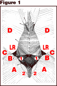
The TLF is composed of a deep and superficial lamina which covers the back muscles from the sacral region through the thoracic region as far as the fascia nuchae.2 Figure 1 depicts the superficial lamina, which shows its connection to the latissimus dorsi, gluteus maximus, part of the external oblique muscle, and the trapezius. The fibers of the superficial lamina attach to the supraspinal ligaments and spinous processes, especially above L4. Caudal to L4, the superficial fascia crosses to the contralateral side and attaches to the sacrum, PSIS and iliac crest.
Figure 1: The superficial lamina. A. Fascia of the gluteus maximus. B. Fascia of the gluteus medius. C. Fascia of external oblique. D. Fascia of latissimus dorsi. Note the cross-hatched appearance of the superficial lamina over the sacrum due to different fiber directions of latissimus dorsi and gluteus maximus. 1. Posterior superior iliac spine. 2. Sacral crest. LR. Part of lateral raphe. Arrows (at left) indicate, from cranial to caudal, the site and direction of the traction (50 N) given to the trapezius, cranial and caudal part of the latissimus dorsi, gluteus medius and gluteus maximus, respectively. Reprinted with permission from: Vleeming A, et al. The posterior layer of the thoracolumbar fascia. Spine 1995;20(7):753.
Due to the different fiber directions of the latissimus dorsi and gluteus maximus, the superficial lamina has a cross-hatched appearance from L4 to S2. At the sacral level, the fibers of the superficial lamina are fused with the fibers of the deep lamina. There is a definite coupling between the gluteus maximus and the contralateral latissimus dors: muscle by way of the posterior layer of the thoracolumbar fascia. Both of these muscles conduct the forces contralaterally during gait and tense the TLF. In so doing, they are important in rotation of the trunk and stabilization of the lower lumbar spine and sacroiliac joint. The deep fascia of the TLF connects to the PSIS, iliac crests and long dorsal SI ligament and encloses the erector spinae muscles. The whole posterior layer of the TLF becomes "inflated" by contraction of the erector spinae.2
Palpation of the fascia over the sacrum where the superficial and deep TLF fuses often reveals tenderness and restrictions in particular directions which require a fascial release to allow the normal transfer of load from, for example, the right lower extremity to the left latissimus dorsi. Any portion of the load transfer mechanism may require a fascial release. In the lumbar region, the deep TLF should be more freely mobile over the back muscles.2 Palpation may reveal hardening and thickening of this tissue, which would require a fascial release. The deep TLF fuses with the fibers of the serratus posterior inferior muscle, which often requires a release.
Vleeming et al.2 recommend training of the gluteus maximus, latissimus dorsi and erector spinae muscles to assist in force closure of the SI joint and stabilization of the lower lumbar area. I feel that besides strengthening of these coupled muscles, it is essential to make sure the fascia is fluid in its motion so that the transfer of load will not be hampered.
References
- Vleeming A, Pool-Goudzwaard AL, Stoeckart R, et al. The posterior layer of the thoracolumbar fascia: its function in load transfer from spine to legs. Spine 1995;20(7):753-758.
- Vleeming A, Mooney V, Snijders C, et al. Movement, Stability and Low Back Pain: The Essential Role of the Pelvis. New York: Churchill Livingstone, 1997.
Click here for previous articles by Warren Hammer, MS, DC, DABCO.


