Part I:"From Guidelines to Practice: What Is the New Benchmark?"
Part II:"Psychosocial Aspects of Low Back Care"
Part III:"Biomechanical Aspects of Low Back Care: The Stabilization System and the Kinetic Chain"
Active care is a proven part of the conservative care armamentarium for neuromusculoskeletal disorders.
Practice-Based Learning
This four-part series is designed to introduce you to a new paradigm of neuromusculoskeletal care. The primary aim has been to provide an opportunity for academic, professional and personal development of each reader. A "best practice" approach integrating the latest developments in active care, the biopsychosocial model, and "evidence-based health care" with existing chiropractic practice has been the major goal of this program.
Innovative approaches to facilitate the learning process have been developed. Traditional passive approaches to learning focus on memorization of didactic material, but it has been my goal to present a critical thinking and problem-solving approach to learning that started with the presentation of the "evidence-based" model of health care (part I of this series). The overall model is termed a "reflective" approach that is practice-based, since its primary aim is to help readers shift their practices to a new model. Our goal is to address:
• the rationale - why;
• the indications - when; and
• the craft - how, for a new skill set for the 21st century chiropractor.
To accomplish this requires a dialectic process by which each reader learns by integrating new skills in practice rather than just passively reading articles or listening to lectures in classrooms. Naturally, this series of articles is just an introduction. To continue the process of change will require attending workshops of this model. Such a practice-based approach is necessarily reflective and experiential, and hopefully will be exciting to each of you embarking on this path.
Aims and Objectives of Reflective Practice-Based Learning:
• evidence-based approach
• critical reasoning skills
• motivation for life long learning
• improved communication/collaboration with other health care professionals
• development and advancement of the profession
• to work at your own pace
• customize implementation of new skills into your practice
• practice workplace as the learning environment.
Tool Box #1 - Driving the Access to Evidence-Based Resources
• United Kingdom Occupational Health Guidelines for Back Pain -http://www.facoccmed.ac.uk - provides an excellent, current reference list of methodologically strong papers
• United Kingdom Health Service health service website - http://www.nice.org.uk- Contains information on "Referral Practice - a guide to appropriate referral-from general to specialist services."
• Chiropractic website which shows how to incorporate "evidence-based" approaches into your practice (http://www.imrci.ac.uk).
• Guide to Assessing Psychosocial Yellow Flags in Acute Low Back . Accident Rehabilitation Compensation Insurance Corporation of New Zealand and National Health Committee 1997. Wellington, NZ. Available from http://www.nhc.govt.nz.
• The Cochrane Collaboration - a leading think tank dedicated to evidence-based health care at http://hiru.mcmaster.ca/cochrane/default.htm.
Functional Goal Setting
Appropriate reassurance and advice may be more important than adjustments and exercise. It is important to mutually establish with the patient that pain relief, reduction of activity intolerances and resumption of physical activities are the key goals. Explaining that hurt does not necessarily equal harm is crucial to success with functional re-activation. It is equally important to avoid disabling forms of advice such as telling a person they have a "ruptured" disc or degenerative arthritis that must be rested. Finally, it is essential to show patients what they can do for themselves.
Tool Box #2 - Driving the Report of Findings
a) Functional Questionnaires (Roland-Morris, Oswestry, Neck Disability Index, Bournemouth Questionnaire);
b) "yellow flags" questionnaire (see Part 2, in DC, Oct. 2 issue);
c) The Back Book for patient education
Screening Evaluation of the Motor Control System
Our goal is to find a dysfunctional pattern that is "linked" to patients' symptoms. To do this, we must evaluate key functions; then we can keep the management prescription simple and fundamental. In this regard, it is wise to remember (from Part 3 in DC, Nov. 30 issue) Kibler's approach, which helps us to see the importance of finding and treating the key dysfunctions responsible for the symptoms.
Table 1 - The Kinetic Chain Approach
History:
• Identify the clinical symptom complex.
Examination:
• Identify the tissue injury complex.
• Identify the source of biomechanical overload.
• Identify the dysfunctional kinetic chain.
Tool Box #3 - Driving the Functional Screening Examination of the Motor System
a) posture and gait
b) respiration
c) stereotypical movement patterns
d) quantifiable physical capacity tests
e) balance and stability tests.
Posture and Gait
This is for orientation and to provide a holistic view of the patient's basic symmetry, global muscle overactivity, and deep muscle inhibition. It requires a little time, but will save time later by focusing the clinician on key areas. Key postural signs that should be sought include the following:
• anterior head carriage
• chin protraction
• scapular winging
• scapular abduction
• shrugged shoulders
• int. rotated arms
• T/L hypertrophy
• ant. pelvic tilt
• lumbar lordosis
• sway back
• flat back
• horizontal grooves in lumbar region
• genu varum
• genu valgum
• genu recurvatum
• mid-foot: pronation or supination
• oblique (unlevel) pelvis
These signs are incredibly useful in practice. For instance, anterior head carriage with chin protraction are positions of overstrain for the upper cervical spine in extension, lower cervical spine in flexion and TMJ. The muscular factors involved are most likely to include inhibition of the deep neck flexors and digastricus, and shortening of the suboccipitals and lateral pterygoids. From these postural signs we will have a picture of what postural cues about which to advise the patient; what muscles to relax and which to facilitate in order to stabilize the TMJ and cervico-cranial joints.
Another very important sign is T/L (thoracolumbar) hypertrophy. This sign of global muscle overactivity is a strong indicator that the deep stabilization muscles in the region are not functioning to protect the individual lumbar joints. Back stability training, such as quadruped single-leg raises may be indicated to teach the patient how to relax the global muscles while performing an exercise targeted for the deep back muscles (see Part 1 in DC, Aug. 6 issue).
Respiration
Faulty respiration is a common source of biomechanical overload of the neck and upper quarter, as well as disturbed abdominal function. Chest breathing overactivates the upper scapular muscles and the scalenes. Failure to use the diaphragm during inhalation can also inhibit normal abdominal activity since they are in a reciprocal relationship. During seated evaluation of relaxed inhalation, look for:
• clavicular elevation
• absence of lateral rib excursion (may need to palpate this)
• paradoxical breathing (p.b.)
• p.b.- supine.
In particular, paradoxical breathing supine in which a person's abdomen goes in during inhalation and out during exhalation is an indication of an uncoordinated pattern.
Stereotypical Movement Patterns
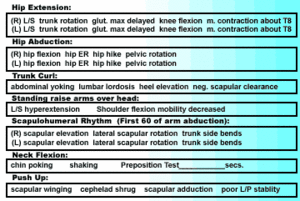
Simple screening tests of specific joint movements can often reveal patterns of joint overstrain and muscle imbalance (overactive and inhibited muscles).
Tool Box #4 - Driving the Evaluation of Muscle Imbalance
• hip extension
• hip abduction
• trunk curl-up
• arm elevation in the sagittal plane (standing arm raise)
• arm elevation in the frontal plane (scapulo-humeral rhythm)
• neck curl-up
• trunk lowering from the push-up.
(Table 1: 7 Exercises and Variations)
These movement patterns are incredibly valuable in practice, since they take only a few seconds per test, and nearly all the major joints are assessed. For instance, the scapulohumeral rhythm test can tell you if your shoulder pain patient needs facilitation methods for their lower trapezius. If in the first 60° of arm abduction the shoulder shrugs, then it is clear that the lower scapular fixators are not balancing the upper shoulder girdle muscles (i.e., upper trapezius and levator scapulae). No amount of medication, injections, soft tissue work, or adjustments are going to correct a rotator cuff problem with impingement supported by excessive superior migration of the head of humerus in the glenoid fossa. Facilitation of the scapular fixators is required.
Quantifiable Physical Capacity Tests
For documentation purposes, and to identify strength or endurance deficits as baselines from which to gauge future progress, it is valuable to use a battery of quantifiable tests. These tests should be safe; have normative data; be simple to perform; not take much time; not require expensive equipment; and be valid. Normative data has been established for tests of squatting, trunk extensor endurance (Sorensen's test), and side bridge endurance (quadratus lumborum and oblique abdominals).
• Squat # of reps (max.50) ______
• Sorensen's ____secs.
• Side Bridge Endurance: (R) ______ secs. (L) ______ secs.
Clinically, these tests are invaluable for patient motivation. If a patient is at 50% or less than the normal for their age and sex, establishing a goal of getting to 85% of normal can be used to give the patient an important target toward which to strive.
Balance and Stability Tests
Balance and stability tests are the final category in the functional screening assessment. They include tests such as single-leg-standing balance and, lunge and reflex gripping of the toes (Vele's test). Many of our musculoskeletal ailments have a key perpetuating factor-disturbed function of the feet. In a patient with knee or ankle pain, it is easy to see the importance of good foot function, but it is also vital to see if one's overall stability is compromised by poor function of the feet. Gait or posture on one leg often amplifies pelvic unleveling, T/L hypertrophy, shrugged shoulders or a head-forward posture. If it does, the feet are just as important as any local findings that may be present.
• Vele's Test: (look for delayed or lack of toe-gripping when leaning forward) _____ side(s)
• 1-leg stance (eyes closed):
(R)_____secs. (L)_____ secs.
pelvic shift______stance leg(s)
pelvic unleveling_____ stance leg(s)
(Norm = 20 sec. with eyes closed < 60 yrs/ 10 sec. > 60 yrs.; Liebenson C. Rehabilitation of the Spine, p. 83)
• Lunge: trunk drift forward ______side
L/S hyperextension ______side
excessive knee shaking ______side(s)
Many patients with poor balance respond to the inclusion of sensory-motor training in their program.
Functional Reactivation
For active care to be practiced, it should be kept simple. Regular progressions should be built-in so patients don't become bored, and the prescription should be linked to activities of daily living and functional end points. Simple reactivation advice is very safe and can begin early in care, with tips about how to modify activities of daily living, such as sitting, bending and carrying. Patient compliance is much better than most would think if functional goals are mutually established and the myth that hurt necessarily equals harm is addressed. The following approaches drive the reactivation of patients.
Tool Box #5 - Driving the Re-activation of Patients
• postural advice
• sensory-motor training
• breathing re-education
• training the deep stabilization muscles
• flexibility training.
a) Postural advice
Kinaesthetic awareness of "neutral" postures is a good starting point for rehabilitation of the motor system. Co-activation of agonist and antagonist muscles is necessary to produce and maintain these aligned postures. First, however, the patient must become aware of how to correct their postural faults such as head- forward posture, protracted chin, shrugged shoulders, swayed back, locked knees, etc.
Simple tools include postural analysis, Brßgger's relief position, abdominal hollowing, cat-camel motions, and chin tucks.
b) Sensory-motor training
Sensory-motor training is one of the best forms of active care, because it is fun for patients, and because results occur so quickly. Simple tools include the single leg balance test, rocker and wobble boards, and balance sandals.
c) Breathing re-education
Faulty breathing leads to cervical, cervico brachial and lumbar problems. Breathing exercises are among the first that are often prescribed and can be used from the first day of an acute episode. Tools include breathing assessment, belly breathing exercises, and rib mobilizations.
d) Training the deep stabilization muscles
Stability training is a key form of safe exercise for patients. It involves learning how to maintain proper form while moving the body against resistance. Specifically, end-range loading of injured joints is limited, especially in the early phases of rehabilitation. Progressions include functional activities, and strength and speed training. Full-range activities are included, so long as agonist-antagonist muscle co-activation is present. Labile tools such as gym balls and balance boards are also utilized, once the patient can demonstrate good motor control. They can increase the isolation of the "deep" muscles.
e) Flexibility training
Flexibility of peripheral muscles is helpful in stabilizing the spine. Stiff ankles, knees and hips will lead to compensatory over stress of the lumbo-pelvic region. However, flexibility training of the spinal muscles has not been shown to prevent injury. In fact, more mobile spines are shown to have a greater likelihood of problems. The tool set for flexibility training includes post-isometric relaxation, active release, myofascial release, reciprocal inhibition, contract-relax, antagonist-contract, hold-relax, and static stretching.
Chart Documentation
Patient progress toward functional goals should be monitored and documented. Simple, clear records are needed so you can stay on top of patient status, progress a program, and consolidate exercises as needed.
Tool Box #6 - Driving the Documentation in Your Charts
a) Roland-Morris, Oswestry, or Bournemouth Questionnaire
b) Neck Disability Index
c) yellow flags
d) quantifiable functional tests
Summary
This last article in a four-part series is designed to summarize the tool sets needed to drive the inclusion of active care into a chiropractic practice. Open-mindedness to change is the first step. Understanding the biopsychosocial model and the biomechanical basis of spine instability is next. Finally, the crafts of a functional screening examination, active care skills, and documentation methods are all needed to help transition a chiropractor from a traditional passive approach to a more active one. The one idea that should be clear as a result of this series is that active care is essential for "benchmarking" chiropractors as experts in managing disorders of the locomotor system.
References
- Liebenson CS, Yeomans SG. Yellow flags: early identification of risk factors of chronicity in acute patients. J Rehabil Outcomes Meas, 4(2), 31-40, 2000.
- Liebenson CS. Motivating pain patients to become more active. Journal of Bodywork and Movement Therapies, 3;143-146, 1999.
- Janda V. Chapter 16: Evaluaiton of muscle imbalance. In: Liebenson C (ed). Rehabilitation of the Spine: A Practitioner's Manual. Williams and Wilkins, Baltimore, 1996.
- Liebenson CS, Chapman S. Lumbar Spine: Making a Rehabilitation Prescription. Williams and Wilkins, 1998 - video.
- Liebenson CS, LeFebvre R, DeFranca C. Cervico-Thoracic Spine: Making a Rehabilitation Prescription. Williams and Wilkins, 1998 - video.
- McGill S, Childs A, Liebenson C. Endurance times for low back stabilization exercises: Clinical targets for testing and training from a normative database. Arch Phys Med Rehabil, 1999;80:941-4.
- Janda V. Chapter 15: sensory-motor training. In: Liebenson C Rehabilitation of the Spine: A Practitioner's Manual. Williams and Wilkins, Baltimore, 1996. 800 638-0672
- Liebenson CS. The safe back workout. JNMS,7(1), 1999.
- McGill S. Low back exercises: Evidence for improving exercise regimens. Phys Ther. 1998;78:754-765.
- McGill SM. Low back exercises: prescription for the healthy back and when recovering from injury. In: Resources Manual for Guidelines for Exercise Testing and Prescription, 3rd ed. Indianapolis, Ind: American College of Sports Medicine. Baltimore, Williams and Wilkins, 1998.
- Richardson CA, Jull GA. Muscle control-pain control. What exercises would you prescribe? Man Ther 1995:1(1):2-10.
- Richardson, Jull, Hodges. Therapeutic Exercise for Spinal Stabilization in Lower Back Pain. Churchill Livingstone, 1999.
- Burton K, Waddell G. Information and advice to patients w/ back pain can have a positive effect. Spine 1999:24;2484-2491.
- McGill S. Stability: from biomechanical concept to chiropractic practice. J Can Chiro Assoc 1999;43:75-87.
- Oslance J, Liebenson C. The Proprio System. 1996. The Gym Ball Store
- Roland M, Waddell G, Moffett JK, Burton K, Main C, Cantrell T. The Back Book. 1996, The Stationary Office, London. ISBN0 11 702078 8 -The Stationary Office.
- Murphy DR, Liebenson C, Ierna GF, Gluck N. Lumbar Spinal Stabilization - Floor Exercises. ACES, 1998. (800) 330-2237.
- Waddell G. The Back Pain Revolution. 1998, Churchill Livingstone, Edinburgh.
- Waddell G, Burton AK, 2000. Occupational Health Guidelines for the Management of Low Back Pain at Work - Evidence Review. Faculty of Occupational Medicine. London.
- Kibler WB, Herring SA, Press JM. Functional Rehabilitation of Sports and Musculoskeletal Injuries. Aspen, 1988.
- Panjabi MM. The stabilizing system of the spine. Part 1. Function, dysfunction, adaptation, and enhancement. J Spinal Disorders 1992;5:383-389.
- Radebold A, Cholewicki J, Panjabi MM, Patel TC. Muscle response pattern to sudden trunk loading in healthy individuals and in patients with chronic low back pain. Spine 2000;25:947-954.
- O'Sullivan P, Twomey L, Allison G. Evaluation of specific stabilizing exercise in the treatment of chronic low back pain with radiologic diagnosis of spondylolysis or spondylolysthesis. Spine 1997; 24:2959-2967.
- Hides JA, Stokes MJ, Saide M, Jull Ga, Cooper DH. Evidence of lumbar multifidus muscle wasting ipsilateral to symptoms in patients with acute/subacute low back pain. Spine 1993; 19(2):165-172.
- Hides JA, Richardson CA, Jull GA. Multifidus muscle recovery is not automatic after resolution of acute, first-episode of low back pain. Spine 1996a;;21(23):2763-2769.
- Bergmark A. Stability of the lumbar spine. Acta Orth Scand Supp 1989; 230(60);20-24.
- Norrs C. Back Stability. Human Kinetics, 2000. (800) 747-4457.
- Linton SJ. The socioeconomic impact of chronic back pain: is anyone benefiting? Editorial. Pain 75:163-168, 1998.
- Morgan D. Concepts in functional training and postural stabilization for the low-back-injured. Top Acute Care Trauma Rehabil 1988;2(4):8-17.
- Lewit K. Manipulative Therapy in Rehabilitation of the Motor System, 3rd edition. London: Butterworths, 1999.
- Liebenson C. (ed). Rehabilitation of the Spine: A Practitioner's Manual. Williams and Wilkins, Baltimore, 1996.
- Yeomans S. Outcomes Management. Appleton & Lange, Stamford, Connecticut, 1999.
- Murphy D. Conservative Management of Cervical Spine Disorders. McGraw Hill, New York, 1999.
Los Angeles, California
Click here for previous articles by Craig Liebenson, DC.


