


The patient was referred for a consultation regarding his consideration for cervical disc surgery and his upper extremity pain. The patient was accepted for treatment for chiropractic care.
History of Present Illness
The patient is a 54-year-old male whose complaint is right anterior shoulder pain, right proximal medial humeral pain, right ulnar groove pain and anterior forearm to right thumb pain. The pain level is rated as an inconvenience and a 5 on a scale of 1-10, with 10 being severe pain with moderate interference in activities of daily living. The quality of the pain is throbbing and burning. He reports numbness at the right lateral aspect of his neck and is intolerant of sleeping on his right side when the pain is at its worst. The patient notes paraesthesias at the right thumb and index finger. Heat therapy improves symptoms.
Physical Examination
Evaluation: Height: 5'7," weight: 210 lbs; vital signs: B/P 140/80 LA; P 88; RR 18.
Inspection: Thickened neck, thick limbs and shoulders, high back; accentuated upper kyphosis of back noted on postural. The patient protrudes head forward and rotates occiput contralateral. He also has an altered posture with cervical anterior carriage with spinal list, increased kyphosis and lumbopelvic anterior carriage.
Musculoskeletal: There is marked tenderness at the forearm flexors, but none to the cervical musculature. There is no facetal tenderness. Distal and proximal pulses are full, regular and strong. No signs for dural tension is exhibited or nuccal rigidity. No Adam's sign (scoliosis) noted. Cervical spine active range of motion is essentially full with pain on extension. Gait is normal and nonassisted.
Orthopedic and Neurological Examination:
| Myotone or Motor Tests | Right | Left |
| C5 Shoulder Abduction | 4/5 | 5/5 |
| C6 Wrist Extension | 4/5 | 5/5 |
Seated deep-tendon reflexes of the upper extremities C5 to C7 demonstrate 2/4 symmetrically. Testing sensory discrimination in bilateral upper extremities C4-T1 dermatomes demonstrates no sensory deficits. Cervical compression is negative for any sprain or radicular patterns. Muscle/myotone testing yields some breakaway quality at forearm flexors, opponens and abductor brevis and longus. Circumferential measurements shows no atrophy.
Record Review and Diagnostic Tests: An MRI without contrast of the cervical spine obtained 6-02-2009 at a regional hospital shows C3-4 osteophyte complex that is degenerative in nature with associated IVF encroachment, C5-C6 right paracentral disc protrusion with desiccated disc, which interprets as degenerative in nature, and severe right foramina encroachment. A T1 hemiangioma is noted.
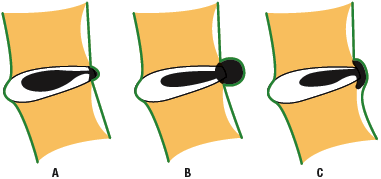
The diagnosis is a right IVF stenosis that is degenerative in nature. This diagnosis, along with the T1 hemiangioma, may contribute to a positional and intermittent right cervical radiculopathy.
Discussion and Recommendations
Disc bulge, facet hypertrophy and flaval ligament thickening frequently combine to cause central spinal stenosis, while degenerative progression with osteophyte bony changes frequently contribute to lateral recess or IVF stenosis. The pain is reproducible on this patient only in the right, lateral recumbent position and on extreme cervical extension. No neurological changes are yielded on examination. Eighty-five to 95 percent of people age 50 have degenerated discs, while 98 percent of those ages 60-80 have degenerated discs. For herniated levels, the most common in the cervical spine is at levels C4-7.
Abnormal findings on MRI do not necessary relate to degree of symptoms. In the case of this patient, the radiologist reads the term protrusion, which signifies contained intact with the posterior longitudinal ligament, and not a noncontained extrusion, which sometimes requires a surgical correction.
I will contact the radiologist at the regional hospital and have him review this MRI again to advise whether the hemiangioma at vertebral level T1 is intradural/extramedullary, within thecal sac but outside the cord, or intramedullary but within the cord. The nature of this can contribute a cervical radiculopathy. In the interim, a detailed discussion with the patient on his condition using anatomical models and drawings commenced.
The patient is still undergoing chiropractic care to date and was cleared on the hemiangioma as noncontributory. He has lost 15 pounds, initiated a weight-training and walking program, is 70 percent pain free and has residual paraesthesias invoking the right thumb and index. He is to repeat his MRI within six months to evaluate progression.
Click here for previous articles by Nancy Martin-Molina, DC, QME, MBA, CCSP.