


Chiropractors have long believed that success and recognition would come by proving the adjustment is more effective than a pill. Much of the profession today is immersed in research and marketing to prove this point.
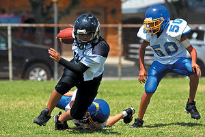 The bigger issue, the overall health and well-being of middle and high school athletes is further compromised by the fact they receive only a medical exam prior to the start of each sports season, having their eyes, ears, nose and throats examined. This exam virtually ignores the critically important musculoskeletal system. This, in spite of the fact that the musculoskeletal system is the system at greatest risk in training and competing in all sports. With many sports being categorized as either contact or collision sports, it is important to remember the body keeps a perfect scorecard of every trauma it endures during one's lifetime. We know youth hides a lot of (biomechanical) sins, so a comprehensive biomechanical exam, producing visual evidence for the examiner and the patient to see, should become part of every young athlete's preparation. There should be no subjectivity or guesswork in this work-up.
Law of Tissue Tolerance
When the loading of a tissue exceeds the capacity of that tissue, compensatory physiological changes occur.
Structural Fingerprint Exam
Excessive and abnormal loading (biomechanical faults) of the musculoskeletal system occurs in all human beings. There is no profession who looks for these faults on examination. There is no profession as qualified as the chiropractic profession to identify and improve these faults, however, too many in our profession are stuck in the near-sighted, reactive system that dominates the care of athletes today. Many allow fear and insurance guidelines to dictate their protocols and recommendations. The very thought that the declining insurance coverage for our active kids will determine the outcome of their health and well-being should give all of us pause for concern.
The first step in incorporating a biomechanical model of care is making the patient understand why a biomechanical exam is important. The Structural Fingerprint Exam is the exam of choice. Even in the absence of symptoms, this exam identifies the biomechanical, or architectural, imbalances, weaknesses, distortion patterns and fixations, which are the predictable factors that will lead to the ultimate wear and tear of a musculoskeletal system. Biomechanical faults are major contributors to almost all sports injuries.
We've found that showing the patient a graphic allows us to explain biomechanical faults in a very succinct and effective manner. As we show the graphic, either as a poster on a wall or in an 8.5" x 11" plastic sleeve, we tell the patient, "All people have biomechanical faults and imbalances that originate in the feet, and produce a compensatory effect up the entire structure. These faults produce abnormal loading in multiple joints, tendons and muscles in the body which are the precursors to sports injuries. Our exam will detect your unique biomechanical faults, which will allow us then to recommend a customized program to proactively address and improve your faults before they become injuries". This explanation is highly effective, despite it's brevity.
The exam begins with a history of what the patient's been through. What sports do they play, what injuries have they had and what tests have they had done? What were the outcomes, what treatments have they attempted and what are the current symptoms they now suffer with.
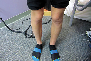 Figure 1: Test for medial arches and alignment of knees
Once the history is complete, I want to know what the patient's goals are. If the goal is merely symptomatic improvement, then I want to answer their goal. I don't eliminate any of the tests I perform on the exam, I only give them a treatment plan with their goal in mind. My experience is that most patients want both symptomatic and corrective care once they visualize the biomechanical faults that are found. This applies to the never-injured and no-current-injury patients as well.
Figure 1: Test for medial arches and alignment of knees
Once the history is complete, I want to know what the patient's goals are. If the goal is merely symptomatic improvement, then I want to answer their goal. I don't eliminate any of the tests I perform on the exam, I only give them a treatment plan with their goal in mind. My experience is that most patients want both symptomatic and corrective care once they visualize the biomechanical faults that are found. This applies to the never-injured and no-current-injury patients as well.
The exam begins with the patient standing, shoes off. The patient looks straight ahead while the examiner palpates the medial arches of the feet along with noticing the (mis)alignment of both knees. As seen in Fig. 1 (increased Q angles bilaterally), there is a gross medial rotation of the knee joints producing significant abnormal loading.
In addition, we perform a digital foot scan (Fig. 2) of the patient in the standing position. This scan provides invaluable information that contributes greatly to the patient's ultimate plan for correction.
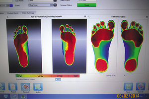 Figure 2: Digital Foot Scan
The exam continues with low back and cervical range of motion, testing for pain and/or restriction. Both findings are highly informative. If there are cervical issues, further compression, distraction and neurological/vascular tests should be performed.
Figure 2: Digital Foot Scan
The exam continues with low back and cervical range of motion, testing for pain and/or restriction. Both findings are highly informative. If there are cervical issues, further compression, distraction and neurological/vascular tests should be performed.
The patient is then tested in the supine position, testing toe-in, straight leg and Patrick-Faberes. Imbalances, pain and restrictions are noted. The patient is then asked to turn over, and in the prone position, lumbar compression, trigger point testing in the quadratus lumborum muscles, gluteus medius muscles and piriformis muscles are performed. A positive test is pain or tenderness upon pressure. The examiner is noting which muscles are sore or painful upon pressure.
The final physical tests are Yeoman's and leg length. Leg length testing is performed differently throughout our profession. The author's recommendation is to measure from a consistent point on the greater trochanter (in the prone position) to a consistent point on the lateral malleolus. The measurement might have to be done several times to get an accurate measurement, but the goal is to determine if there is an anatomical short leg. If so, this requires a lift as part of the correction.
The final test is biomechanical x-rays. These are mandatory, and this separates us from all other professions. Just as the medic requires blood and urine tests, we must become experts on the wealth of biomechanical information that is only provided with standing x-rays. We take four views, two of the cervical spine and two of the lumbo-sacral spine, both series in the standing position and barefoot. The views are A-P and lateral, with the cervical x-ray being an open mouth view/lower cervical.
Once all of these tests are performed, a report of findings is set up to show the patient their unique biomechanical faults and what programs are available to address these faults. The biomechanical information on x-rays and the report of findings will be addressed in future articles in this series.
The world of sports has grown to immeasurable heights in our society today. There are more kid's groups, from soccer to football to baseball to tae-kwon-do, and each and every child will suffer needless injuries and premature degeneration if we continue with the model that is used today. My mission is to help lead our profession, with the application of the biomechanical model, by changing how these young athletes are cared for. This change could dramatically affect their long-term quality of life as well as provide relief to the out-of-control, escalating healthcare costs of today.
Editor's Note: This is part two of a six part series by Dr. Maggs. To see part one, visit www.dcpracticeinsights.com.
Dr. Tim Maggs has been in private practice for 35 years and is the developer of The Structural Management® Program and The Concerned Parents of Young Athlete's® Program. He travels the country teaching his program Biomechanics, Imaging and Care of the High School Athlete. For more information, visit www.CPOYA.com.