


As we noted in our previous article, with a positive Derifield (+D),1-2 the doctor observes the reactive (shorter) leg in the prone position that becomes longer or "crosses over" in the flexed position.
A negative Derifield1-2 is indicated when the doctor observes a shortening of the reactive (shorter) leg in the prone position that becomes shorter, stays short or fails to cross over in the flexed position. The negative Derifield test result suggests a θY-axis segmental dysfunction manifesting as rotation of the ilium or sacrum relative to the longitudinal axis of the body. Four different listings of sacroiliac joint dysfunction may be indicated by a negative Derifield test: internal (IN) ilium, external (EX) ilium, posterior rotation of the sacral base on the left or right (P-L or P-R) and anterior rotation of the sacral base on the left or right (A-L or A-R.) In this discussion, we address two of the additional listings: EX ilium and posterior rotation of the sacral base (P-L or P-R.)
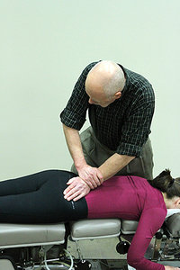 Fig. 1: Right SI fluid motion test, performed to determine sacroiliac fixation.
Differentiation: EX Ilium, P-L / P-R
Fig. 1: Right SI fluid motion test, performed to determine sacroiliac fixation.
Differentiation: EX Ilium, P-L / P-R
To differentiate among the possible outcomes of a negative Derifield test, use the θY-axis segmental dysfunction test for the pelvis.3 Stand at the level of the patient's thigh facing obliquely inferior on the side to be tested. Instruct the patient to lock the knee on the side tested. Help the patient to extend the lower extremity at the hip to its end range or point of resistance. Finally, passively extend the hip a few degrees into resistance to engage the sacroiliac joint on the side of involvement. Observe changes in the length of the reactive leg in the prone extended position. A positive test is suggested if the reactive leg stays short in the extended prone position.
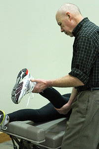 Fig. 2: Right θY-axis segmental dysfunction test for the pelvis. This is an isolation test performed
immediately following a negative Derifield leg check finding (-D). The purpose of the test is to narrow down the possible θY-axis pelvic misalignments from four to two possibilities.
Next, flex the knees and observe for changes in the leg on the side tested. If the leg becomes long in the flexed position, the test suggests EX ilium or posterior rotation of the sacrum on the involved side. Differentiate using an articular pressure test,3, 5-6 to determine the appropriate listing and correct line of correction. Posterior rotation (P-R/ P-L) of the sacrum tends to occur more frequently than an EX ilium. A positive articular pressure test is indicated when the legs become even in the prone extended and flexed positions.
To perform an articular pressure test for a posterior rotation of the sacrum on the side tested, stand on the side opposite the involved sacroiliac joint. Contact the sacral ala lateral to the first and second sacral tubercle using the hypothenar or pinky side of the heel of the hand. Apply a firm medial-to-lateral and posterior-to-anterior pressure parallel to the sacroiliac joint space. If the legs become even in the extended prone position and even in the flexed position, the test is positive for a posterior rotation of the sacrum.
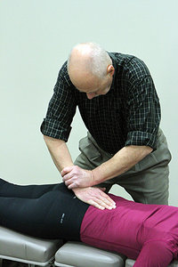 Fig. 3: Adjusting setup for right posterior rotation of the sacrum (P-R).
Adjusting Techniques
Fig. 3: Adjusting setup for right posterior rotation of the sacrum (P-R).
Adjusting Techniques
To adjust a posterior sacrum, stand on the side opposite the joint involvement. Contact the sacral ala lateral to the first and second sacral tubercle using the hypothenar eminence of the hand. Apply a thrust from medial-to-lateral and posterior-to-anterior parallel to the sacroiliac joint space.7 (Note: The line of correction for the thrust is identical to the direction used in the articular pressure test, discussed in part 1 of this article.)
Following the adjustment, perform another Derifield leg check. Legs should be even in the prone extended position and even in the knee-flexed position.1-3 Then test the sacroiliac joint for mobility by repeating the SI fluid motion test. The joint will now typically be freely movable on testing.
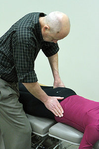 Fig. 4: Adjusting setup for right EX ilium.
If the legs do not become even after the articular pressure test for posterior rotation of the sacrum, test for an external (EX) ilium on the involved side. To test for an EX ilium, stand on the side of involvement facing obliquely superior. Contact the inferior lateral aspect of the PSIS with the heel of the hand, and apply a gentle, but firm pressure from lateral-to-medial and from inferior-to-superior parallel to the sacroiliac joint space. (Recall that an ilium rides inferior or down the articulation with an EX misalignment.) If the legs become even in the extended prone position and in the flexed position, the test is positive for an EX ilium on the side tested.
Fig. 4: Adjusting setup for right EX ilium.
If the legs do not become even after the articular pressure test for posterior rotation of the sacrum, test for an external (EX) ilium on the involved side. To test for an EX ilium, stand on the side of involvement facing obliquely superior. Contact the inferior lateral aspect of the PSIS with the heel of the hand, and apply a gentle, but firm pressure from lateral-to-medial and from inferior-to-superior parallel to the sacroiliac joint space. (Recall that an ilium rides inferior or down the articulation with an EX misalignment.) If the legs become even in the extended prone position and in the flexed position, the test is positive for an EX ilium on the side tested.
To adjust an EX ilium, stand on the side of the joint involvement. Contact the inferior lateral aspect of the PSIS with the proximal thenar of the hand, and apply an arcing thrust lateral-to-medial, posterior-to-anterior, and inferior-to-superior parallel to the plane line of the sacroiliac joint.7 (Note: Again, the line of correction for the thrust is identical to the direction used for the articular pressure test.)
Following the adjustment, perform another Derifield leg check. Legs should be even in the prone extended position and even in the knee-flexed position. Then test the sacroiliac joint for mobility by repeating the SI fluid motion test. The joint will now typically be freely movable on testing.
References
- Laufenberg PC. Thompson Terminal Point Handbook. Davenport, IA: J Clay Thompson, 1974.
- Thompson Educational Workshops. The Thompson Technique Reference Manual. Elgin, IL: 1984.
- Fuhr AW, et al. Activator Methods Chiropractic Technique, 2nd Edition. St. Louis: Mosby-Elsevier, 2009.
- Kasperbauer R, et al. "Gonstead Methodol-ogy Institute: Case Management." Continuing Education, Logan College of Chiropractic, January 2014.
- Haas M, Peterson D, Panzer D, et al. Reactivity of leg alignment to articular pressure testing: evaluation of a diagnostic test using a randomized crossover clinical trial approach. J Manip Physiol Ther, 1993;16(4):220-7.
- Khauv KB, John C. Health-related quality of the improvements in adult patients with chronic low back pain under low-force chiropractic care: a practiced-based study. Chiropr J Aust, 2011 Dec;41(4):118-122.
- Pettersson, HA, Green, JR. How to Find a Subluxation. Davenport, IA: Black Athena Press, 2003.
Authors' Note: Part 3 of this article will address the remaining two listings: IN ilium and anterior rotation of the sacral base (A-L / A-R).
Dr. Howard Pettersson, a 1976 graduate of Logan College of Chiropractic, is an associate professor of technique at Palmer College of Chiropractic. He was the senior editor of Activator Methods Chiropractic Technique – College Edition, published in 1989, and published Pelvic Drop Table Adjusting Technique in 1999. His most recent publication, written with Dr. Green, is How to Find a Subluxation, published in 2003.
Dr. J.R. Green is a 1988 Graduate of Palmer College of Chiropractic. He retired from the Palmer faculty after many years of teaching basic sciences and chiropractic technique. He is currently in private practice in Galva, Ill., and is also an adjunct professor of chemistry with the Eastern Iowa Community College District. Dr. Green was one of the writers of Activator Methods Chiropractic Technique (1997) and also worked as a technical writing consultant on Activator Methods Chiropractic Technique – College Edition and Pelvic Drop Table Adjusting Technique.