


Dual-energy X-ray absorptiometry (DXA) is still the gold standard for measuring bone mineral density. This test is used to assess fracture risk, especially in postmenopausal women. The World Health Organization defines osteoporosis as a BMD T-score of -2.5 or lower at any location, or having a previous fragility fracture.
There is even less of a consensus on the definition of osteoporosis in men. The WHO recommends similar T-score thresholds in men ages 50 and older. This is a problem because men generally have a larger skeletal structure and the fracture risk is less than half of women starting at age 55.3
 Even though individuals with a T-score below the -2.5 cutoff are considered at higher risk of fracture, they do not account for the majority of fracture cases in either women or men, according to the Rotterdam Study.4
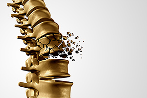
Is the "Gold Standard" Enough?
The DXA scan generates a two-dimensional image of a complex three-dimensional structure and reports a quotient of the bone mineral content divided by the bone area. The obvious problem with this method is that larger bone will be determined to be superior in strength, but may actually have the same bone density as smaller bone.5
Mineral content does not necessarily determine bone strength.6 The overall strength of a bone depends on the proportion of the cortical and trabecular tissues, their morphologies and material properties, and the interactions among these traits. There is also the role that the individual's unique genetic makeup contributes to bone strength.6 Furthermore, technical factors in patient positioning, physical artifacts, and degenerative joint and disc disease with exuberant osteophytic formation, aortic calcification and surgical clips are a few examples in which the results of the DXA score can be falsely elevated.7
There are other methods for measuring BMD, but most are complementary to and dependent on the DXA scan. The first and most common tool is the FRAX scoring system, which is a clinical questionnaire. It is widely used to assess risk of fracture over a 10-year period. FRAX can be used with or without a DXA score, but is frequently used as a complement (www.sheffield.ac.uk/FRAX/tool.aspx?country=9).
The Trabecular bone score (TBS) is an analytical tool the that performs novel grey-level texture measurements on the DXA images, thereby capturing information relating to trabecular microarchitecture.8 The TBS can also be included in the FRAX scoring system. The problem with these tools is that they both rely on the accuracy of the DXA scan.
Quantitative CT (QCT), which can provide 3-D volumetric measurements using a low-dose scan protocol, generates reasonable estimates of strength through engineering-based analyses such as finite element analysis (FEA): robust computer programing. QCT is used mainly for evaluating the hip and lumbar spine. It is less accurate in the thoracic region.
Another variation of QCT is high-resolution peripheral quantitative computed tomography (HR-pQCT), which is primarily used to assess tibia and radius bone architecture and density. The correlation between QCT and other non-vertebral osteoporotic fracture is not clearly delineated; higher exposure to ionizing radiation and cost make this modality less attractive as a screening test. Additionally, there are problems with which methods are needed to adjust for variation in marrow fat and soft-tissue density.9
Quantitative ultrasound (QUS) has been used to evaluate bone structure and fragility, but this procedure has been done primarily on the calcaneus; and the correlation between bone structure in the heel and spine has issues depending on the bias of the researcher. QUS cannot be used as a stand-alone method for the diagnosis of osteoporosis because it is operator dependent, difficult to reproduce and only provides an indirect measurement of bone density.
Bone turnover markers (BTMs), which are released during bone remodeling, can be assessed, but there is insufficient data and understanding of bone turnover markers for the diagnosis of osteoporosis. It is not able to be used even to identify candidates for treatment; or for the determination of the length of time to pause bisphosphonate treatment. More research needs to be done to understand the behavior of BTMs before we can confidently rely on these markers.
The experts have only recently endorsed the use of BTMs, especially serum C-terminal telopeptide of type 1 collagen (CTX) and serum procollagen type 1 N propeptide (P1NP), as short-term monitoring tools to help clinicians assess the responses to osteoporosis therapies and appropriately adjust treatment regimens before waiting for the next DXA scan. The data is very promising: https://doi.org/10.1016/j.jocd.2019.03.004; just not quite ready for primetime.10
High-resolution MRI is another technique available for bone assessment, with the advantage of being ionizing radiation free. To obtain micro-architectural data of trabecular bone, MRI is used to image the peripheral skeleton (distal radius and calcaneus), but progress is being made using it on other skeletal regions like the proximal femur.11 So far, the use of MRI to evaluate vertebral fractures has been limited to differentiating benign or traumatic fractures from pathological ones.
Studies using 7 T MRI of the femur can detect bone microarchitectural deterioration in women with fragility fractures whose BMD is the same as those who don't have any fractures.12 But there are not many 7 Tesla units available and the scan times are long. Of course, study is expensive, but the microarchitectural parameters might be used as an additional tool to detect patients with poor bone quality who cannot be detected by dual-energy X-ray absorptiometry. This is a promising technique, but again, more research needs to be done.
What's Missing: Identifying Patients Who Don't Require Any Medication
The DXA scan is a great method for screening patients, but we really need another method for determining categories: patients who truly need pharmaceutical intervention and those who would benefit from lifestyle changes without needing medication. We also need to have better tools for following up with patients on medication. For example, how many patients on medication only get a DXA every two to five years? Right now, it is difficult to determine when to stop or pause the medication, or even if the medication is doing the patient any good.
Medications for osteoporosis vary in mechanism of action: anti-resorptive, which are the bisphosphonates and denosumab, primarily increase endocortical bone, bolstering mineralization by decreasing bone resorption. Anabolic medications (teriparatide and abaloparatide) stimulate periosteal and endosteal bone formation without large changes in cortical thickness.
Because of the differences in the mechanisms of these various drugs, it would be beneficial to select a treatment based on a patient's unique bone structure and pattern of bone loss; using a more targeted approach to treat each patient according to their own unique skeletal characteristics. There are attempts to do this, but it seems to me from my review of the literature we have not be able to achieve this goal. We still need more research. The treatment of uncomplicated postmenopausal osteoporosis is still a work in progress.
Editor's Note: Part 2 of this article (December issue) discusses chiropractic interventions that support diagnostic findings and can help manage osteoporotic patients. Complete references supporting the citations in both parts accompany the digital version of part 2.
Click here for more information about Deborah Pate, DC, DACBR.