


This article, and the next, focuses on the lateral side of the sacroiliac (SI) joint, which I refer to as the "ilio-sacral" (IS) joint, to remind you of the focus on the ilium. The nomenclature here is the standard of the osteopathic methods of muscle energy. Low-force adjusting is especially critical for unstable and hypermobile SI joints. My first introduction to the world of low-force, osteopathic-style correction was through my failures. I observed patients I couldn't help, but whose back pain was solved by these methods.
In assessing any subluxation, we look for tenderness and restriction. Tenderness is a challenging indicator for SI problems. The posterior SI joint ligaments may or may not be tender. The usual tender areas in an SI problem include the lateral surface of the ilium, palpated from lateral to medial, directly lateral to the SI joint. You have to slide two or three inches lateral to the posterior superior iliac spine (PSIS) to get enough tissue slack, then push directly medial to assess for tenderness. Another tender site is the posterior-lateral border of the sacrum itself, usually at one specific level. The SI joint is long, so you'll need to test at least three levels along the sacrum and the ilium, superior to inferior.
The initial palpation test should assess general or global stiffness. Remember the principles of initial response testing (www.chiroweb.com/archives/19/16/12.html), assessing at the beginning of the motion, rather than moving the joint through its whole range. For the IS joint, we'll attempt to glide it along its plane of motion.
Here are four tests, two supine and two prone. First, with the patient prone, contact the ilium just lateral to the sacrum, and push it in an anterior lateral direction, along the joint line. Second, reach around to the front, grab the anterior superior iliac spine (ASIS) with your curved fingers, stabilize the sacrum with your other hand, and test for an anterior-to-posterior motion, again along the joint line. The third test is supine. Push the ASIS in a posterior medial direction along the joint line. Fourth (also supine), reach around to the back, grab the PSIS and pull it anterior and lateral. For any of these motion tests, the keys are specificity of direction and the right amount of force. If you are not directly along the plane of the joint, you will not feel the motion. Too little force and nothing moves; too much, and you'll somehow also miss the motion. I personally like these direct tests of motion more than the commonly taught "marching" test.
The SI joint often becomes hypermobile. These same tests can help you find too much motion or laxity. If your impression is one of "slop," or laxity, you'll do the test in a slightly different manner. Take the joint through the whole range of motion, rather than assessing just the beginning feel. The challenging aspect of hypermobility is that the muscles all around the joint tighten up to attempt to stabilize the joint, so you can be fooled by the muscular tightness and stiffness. You have to isolate the joint to find the hypermobility, which usually resolves when the restricted areas in the SI (and all around the region) are corrected. I'll often use an SI or trochanter belt when there has been trauma; when a sacro-occipital technique (SOT) category 2 shows up; or when I can palpate hypermobility. Another excellent test utilizes a belt: Have the patient bend forward, so you can assess range of motion and discomfort; then put a belt on the patient, and have him or her bend again. Is it easier to bend with the belt on? If so, a belt is needed for at least six weeks. When the patient is done with the belt, I advise him or her to store it in the glove compartment of the car, and use it just on long drives, long sits, or during physical labor.
In complicated cases, I'll assess and correct, at minimum, the lumbar spine and the lower extremity, and use visceral manipulation on restricted areas in the lower abdomen. Proper exercise rehabilitation also is critical - especially the avoidance of "one-leg-standing" exercises. Certain patients seem to get better, but easily relapse, either from physical labor or extensive sitting. Such patients can be good candidates for proliferant injections, a medical procedure that helps tighten up the lax ligaments.
Upslips and Downslips: Ilio-Sacral Shears
I want to introduce to you an IS pattern with which most of you probably are unfamiliar. The ilium can shear vertically on the sacrum; this is called an upslip or downslip. When this occurs is critical, because shears are nonphysiological patterns, rather than rotations within the joint's normal planes of motion. In this shear, the ilium has ridden superior or inferior on the sacrum, losing its normal relationship. Once you know how to assess for IS shears, they become obvious, and relatively easy to correct.
What causes these shears? They are almost always traumatic, although the patient doesn't always remember the trauma. Any fall on the buttocks can cause an upslip, as can a motor vehicle accident, particularly when the driver stomps on the brake just before impact, sending a shock wave up the leg. They can also happen from a trauma as simple as stepping off an unexpected step. Downslips can occur iatrogenically, from having the leg pulled in a forceful manner. They can occur when a person gets his or her foot stuck in a hole or in mud, and has to pull to get the leg freed. Imagine getting one leg stuck in quicksand, and what happens to the pelvis as you are pulled out. Shears are more common in pregnancy or childbirth, or in anyone who has lax ligaments.
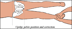
How is an upslip diagnosed? In an upslip, the IS joint on the side that has sheared upward is quite restricted. Besides the global palpation test mentioned earlier, I also test the ilium's inferior glide. Push inferior on the top of the iliac crest, either prone or supine. The muscles around the joint will also be quite tight, compensating for the ligamentous strain pattern. The key for this subluxation is bony positioning. This is a true joint displacement, and a restriction. With the patient supine, palpate the ASIS, the iliac crest, and the pubic symphysis; they will all be cephalad. Check the leg length; it will be substantially shorter on the upslip side. In the prone position, the PSIS and the ischial tuberosity palpates will be cephalad. All of the structures of the pelvis will be cephalad, unlike a sagittal rotation pattern, in which the ASIS and PSIS go in the opposite directions. The combination of the unique positional indicators with joint restriction on the cephalad side indicates the upslip.
The downslip is just the opposite, but it's easy to be confused with the upslip. Is the right side an upslip, or is the left side a downslip? The side of restriction reveals which side is involved. In the downslip, all of the landmarks are caudad, and that side is restricted. The specific restriction is a lack of superior glide. Test cephalad motion at the ischial tuberosity, or with a broad contact on the lateral surface of the ilium. With the patient supine, here's what you'll find on the side of restriction: the iliac crest will be caudad, as will the ASIS and pubic symphysis, and the involved leg will be long. With the patient prone, the PSIS and the ischial tuberosity also will be caudad.
How do we correct the shear? In an upslip, the goal is to pull the ilium inferior. Do this with a thrust, pulling on the leg at the ankle. Ideally, do this prone, but it can also be done supine. Either way, begin by abducting the hip slightly to get relaxation. Start with a slow pull, with the leg internally rotated to lock the hip. Traction up through the kinetic chain, until you feel the pull through the SI joint. Have the patient cough. During the cough, pull inferior, releasing the joint. You may notice an audible release, but don't be attached to this. Go back and recheck your positional and motion indicators.
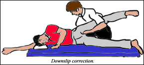
The downslip correction is more cumbersome. In a right downslip, have the patient lie on his or her left side, and support the leg in an abducted position. Your contacts, using both hands, are on the ischial tuberosity and the ilium itself. Bend your knees and lift the patient's leg toward the ceiling for 15-30 seconds, to release the area. Continue applying pressure, and push the cephalad until you feel the joint release. (This feels somewhat like pushing a boat through mud. If you use too little or too much pressure - nothing happens. With just enough pressure, the boat begins to slowly move. That's the feeling you're looking for.) Once you feel the joint release and move, recheck your indicators.
As many of the great chiropractic teachers have emphasized over and over, SI joint integrity is critical. SOT has been a real leader in our profession in its understanding of the importance of low-force correction of this unstable area. My next article will complete the IS chapter, with a look at PI and AS ilia, iliac internal and external rotations (flares), and more on the pubic symphysis. For now, I hope I've given you some new ideas.
Resources
- George Such. Synthesis Seminars, Breitenbush, OR, 1998.
- Rex L. Introduction of muscle energy techniques, (course notes), Ursa Foundation. Edmonds, Wa. 1996. pp. 244-249.
- Greenman P. Principles of Manual Medicine, 2nd edition. Williams and Wilkins, 1996.
- Chauffour P. Mechanical Link 1 (seminar). San Francisco, Ca 1997.
- Heller M. Framework 1, Low Back and Pelvis (seminar). Ashland, OR, 2002.
Click here for more information about Marc Heller, DC.