


Real-time sonography and the use of high-frequency, high-resolution transducers allow for high-quality images of soft tissues to be obtained. Ultrasound imaging has primary importance and wide acceptance in cardiology, gastroenterology and obstetrics, and in several surgical diagnostic procedures. One major advantage of ultrasound compared with other medical imaging modalities is real-time scanning of moving and static soft-tissue structures with reasonable grayscale contrast. The major disadvantages are relative poor spatial image resolution and the fact that because ultrasound does not pass bone, several soft tissues cannot be imaged routinely.
I am aware of several articles that indicate ultrasound can be utilized to visualize ligaments and tendons in the adult spine. I must state that current information does not support this opinion. Even if the technology had improved to the point of making this imaging modality useful for the adult spine, other modalities, such as magnetic resonance (MR) and computed tomography (CT) imaging, remain the gold standard for imaging the spine, and I sincerely doubt this will change anytime soon.
There are many occasions in which ultrasound can be useful and cost-effective for imaging other musculoskeletal regions. The wrist, elbow, knee, ankle, fingers and shoulder can be examined quickly and with less patient discomfort than more invasive procedures, and are less expensive to examine with ultrasound than with other imaging modalities. Trauma to tendons, cysts/fluid collections, neuromas and foreign bodies can be diagnosed and evaluated.
Tendon imaging can be performed with ultrasonography and MR. Both imaging modalities have been improved in recent years because of technical advancements and a better understanding of tendon pathology. For example, in many cases, tendon rupture seems to be the final stage of tendonitis; ultrasound can be used to predict the risk of tendon rupture.
The main disadvantage of ultrasound involves the difficulty in documenting the images in a standardized manner, and therefore, in gaining exactly reproducible diagnoses. This is where MR is superior - not to mention that the spatial resolution is far superior. However, an ultrasound examination may be useful when following the progress - or lack thereof - in the treatment of the tendon abnormality. Repeating an MR study can be expensive, but repeating several ultrasound studies is not unreasonable, particularly once the diagnosis has been confirmed by MR.
Let's review the imaging of tendons by way of ultrasound. Tendons represent a portion of a muscle, and consist of collagen fibers that transmit muscle tension to a mobile part of the body. The fibers are bound together in a three-dimensional network of endothelium septa called the peritendinae. They originate from a fine connective sheath, the epitenon, which surrounds the entire tendon. In large tendons, blood and lymphatic vessels, together with nerves, run within these septa, while small tendons are almost avascular. Tendon sheaths completely or partially cover a portion of the tendon, where it passes through fascial slings or osseofibrous tunnels. They promote gliding and contribute to the nutrition of the tendon tissue. A tendon sheath consists of an outer and inner layer, which are intimately attached to the tendon. Both may be connected by a mesotenon. Long tendons, such as the patellar or the Achilles, are not covered by a synovial tendon sheath, but lie within highly vascularized loose areolar and adipose tissue, the paretenon.
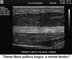
Sonographically, tendons appear as fibrillar structures with hypoechoic (resulting in a bright tissue appearance) collagen fibers and hyperechoic (resulting in a dark tissue appearance) endotendineal septa intermingled between them. The number of fibrils visible with sonography correlates closely with the frequency of the ultrasound probe used. With MRI, normal tendons appear as homogenous hypoechoic structures; therefore, ultrasound can be used to evaluate the tendon in a more dynamic manner, once the location of an injury has been identified.
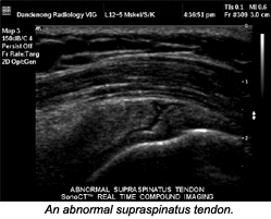
On sonograms, the distinctive signs of tendonitis are hypoechoic foci, with or without tendon thickening. With high-resolution imaging, the lesions appear oval with blurred borders, and seem to be orientated parallel to the course of the tendon fibers. In later stages, generalized thickening with widening of the distance between fascicles is present. Abnormalities of the tendon sheath are, in most cases, clearly visible in the form of hypoechoic thickening of the sheath, due to fluid accumulation.
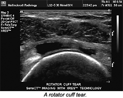
Signs of fresh tendon rupture are similarly visible with sonography or MR, at least in principle. When using ultrasound, characteristic findings, such as thinning or disruption of fibers, may be masked by an overlying hypoechoic hematoma or artifacts. With sonography, partial tears principally are visible in the form of circumscribed hypoechoic lesions. Ultrasound and MR may be used jointly to diagnose tendon diseases, and for clinical follow-up.
The following is a brief list of the more common locations/conditions for which ultrasound can be used, either as a diagnostic modality alone or as an adjunctive modality to MR for diagnosis and clinical follow-up:
Groin: Ultrasound can be used in the management of chronic groin pain, a common problem in sports that is often difficult to diagnose and potentially career-ending for the athlete. Ultrasound is an alternative to contrast herniography for the diagnosis of impalpable inguinal and femoral hernias, providing comparable results while avoiding the need for injection and allowing other structures to be assessed simultaneously.
Knee: Ultrasound is extremely good at characterizing tendonitis and diagnosing various lumps and bumps, such as synovial or meniscal cysts. Unfortunately, many of the important knee injuries that occur in sports are not well-suited to examination by ultrasound. MR is clearly superior in the assessment of meniscal tears, cruciate lesions and osteochondral lesions.
Foot: Ultrasound is most often used to assess tendonitis. However, two recent developments that have relevance to sports concern Morton's neuroma and posterior ankle impingement.
Wrist: One of the more common disorders that may be evaluated with ultrasound is carpal tunnel syndrome (CTS). The etiology of CTS is primarily the encroachment on the median nerve, due to either a decrease in size of the tunnel (mainly osseous causes), or an increase in the volume within the confined space of the tunnel.
In the transverse imaging plane, the ulnar artery is the medial landmark of the carpal tunnel. Imaging must be performed with the scan head in a plane perpendicular to the tendon surface, to eliminate the anisotropic effect. The tunnel contains the flexor digitorum tendons, which are hyperechoic. Anterior to the tendon is the median nerve. The median nerve has a characteristic appearance that differentiates it from the fibrillar hyperechoic tendons. The nerve is hypoechoic with a hyperechoic border and shows multiple bright reflectors in the transverse imaging plane. The median nerve is rounded or oval in the proximal wrist and flattens progressively as it courses through the carpal tunnel. Within the tunnel, the nerve is in intimate contact with the flexor retinaculum; its size remains constant, but its shape is quite variable.
In the longitudinal imaging plane, the nerve is demonstrated coursing parallel and superficial to the flexor digitorum tendons. The sonographic appearance of the nerve in this plane demonstrates hyperechoic continuous anterior and posterior borders (the nerve sheath), and is distinguished easily from the characteristic fibrillar-appearing tendons that lie posteriorly.1 Ultrasound provides a painless, convenient method of following the progress of disease in such cases. Other conditions in the hand that can be evaluated with ultrasound are foreign bodies, tendon rupture, ganglion cysts and synovial cysts.
Comparison MR
Musculoskeletal ultrasound is still an emerging subspecialty, made possible by recent advances in technology. State-of-the-art ultrasound systems can resolve exquisite superficial soft-tissue detail. Equipment that can effectively suppress image noise while operating at a wide dynamic range and ultra-high transmit frequencies also is important, as it allows for the demonstration of subtle variations in tissue texture and the differentiation of anechoic fluid from markedly hypoechoic tissue.
Ultrasound costs less to perform than MR, and even more importantly, it is interactive. Unlike MR, the ultrasound examiner is in the room with the patient, probing the affected part with the transducer and directly correlating the site of reported pain or tenderness with its scan appearances.
However, ultrasound is an operator-dependent modality. Poor results generally indicate poor technique, rather than a poor test, and it is the limited availability of good training that limits ultrasound growth. Remarkable technological developments are ongoing, and many frontiers remain to be explored.
In imaging the hand and wrist, ultrasound can often be more convincing than MR, because routine comparison views of the normal side provide compelling objectivity. Carpal ligament injuries can be readily appreciated by ultrasound, but are typically missed on MR, because the signal obscures adjacent joint space fluid.
MR is the more appropriate modality for defining pathology, and definitely should be utilized when ultrasound yields equivocal findings.
References
- Related case study. www.wipro-ge.com/ultrasound/msucts.html.
- I continue to be indebted to Philips Medical Systems - online at www4.medical.philips.com:80/Education/education.asp - for permission to use its images.
Deborah Pate, DC, DACBR
San Diego, California
![]()
Click here for more information about Deborah Pate, DC, DACBR.