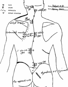
A 60-year-old, retired, white female comes in on the kind referral of her massage therapist. She had a complaint of left mid-thoracic spinal and trunk pain of about a year's duration, and left cervicothoracic pain that developed more recently.
She reports many stressful events over the past few years, with her husband's protracted mental debilitation and recent death. She suffered a fall onto her buttocks on ice about 10 years ago, but reports experiencing little pain after the incident, with no apparent residual effects. She otherwise remembers no significant musculoskeletal trauma other than "normal kid stuff." She had a heart attack at age 34 but seems to have made good recovery and reports no blockage of arteries on a recent angiogram. She takes Tylenol for her back pain, but this is not very effective. She had a hysterectomy at age 49, following a diagnosis of cervical cancer.
Reviewing her standing lumbopelvic X-rays reveals the appearance of a torsioned pelvis with clockwise rotary displacement (cephalid view). There is a left tilt of the sacrum, with the coccyx sitting left of the midline and significant declination of the right sacral base. The left iliac crest appears higher than the right. I see a sweeping left rotary curve in the thoraco-lumbar region. I see no evidence of costal luxation or subluxation on the thoracic A-P view. She has some mild lipping and spurring in the thoracic and lumbar vertebral bodies, and other degenerative change, commensurate with her age. Otherwise, her X-rays appear normal, and there is no suggestion of leg-length inequality.
On visual exam with her in open-back gown, I see that she has good development and alignment in the lower extremities with equal muscle mass. Her panty line declines to the right. There is mild right apex curve at the lumbar region and the right flank fold is very prominent compared to the left. Above this, the lower thoracic spine sweeps left, and in the mid-thoracic region, sweeps back right. Her left shoulder is elevated compared to the right, and her left ear is lower than the right. The left scapula is mildly winged. Palpating the iliac crests with her standing reveals a low right crest. Sitting leg raise, combined with neck flexion and Valsalva maneuver, does not produce spinal or extremity pain or peripheral dysesthesia.
On sitting and prone palpation, I find two areas of biomechanical distress with localized guarding spasm in the cervical spine in opposite rotation: right lower and left upper. The thoracic spine exhibits fixation and loss of springy motion at the outer apices of the rotary curves: left upper, right middle and left lower thoracic rotation. The lumbar is rotated clockwise with fixation at the L3/4 level. Thoracic and lumbar segmental dysfunctions exhibit reflex myospasm. The left intercostal regions at T4/5/6 are painful to probing. The sacroiliac joints are not painful to hard probing, even though they are asymmetrical, and the lumbosacral region is not painful to hard P-A compression. The right sacrotuberous ligament is very hypertonic compared to the left. All visual and palpatory findings are illustrated and charted above. Supine leg check after knee-to-chest flexion reveals legs of apparent equal length.
Discussion: My impression is that this patient is experiencing chronic intercostal neuralgia in the left mid-thoracic region that is related to the spinal biomechanical lesions at the T4/5/6 levels. Beyond this, I believe she may have experienced a fall in her youth that caused displacement of the sacrum and resulted in rotary compensation of the developing spine above. I have observed this rotation in compensatory opposition above a tilted and unleveled sacral base before. There are chronic biomechanical impairments at the apices of all rotary curves. The very taut sacrotuberous ligament in the right gluteal region seems to be another characteristic of this syndrome. This configuration may have developed after the fall on ice some 10 years ago, but I doubt it. Such an injury would have been very symptomatic in adulthood. Also, the sacroiliac and lumbosacral regions show no present signs of distress or inflammation. It is much more likely that she experienced a fall before bony maturation, with permanent remodeling of the sacroiliac joints.
Today, we discuss the nature of her condition and the need for intense, anti-inflammatory cryotherapy and other anti-inflammatory measures to the left mid-thoracic region. We apply 10 minutes of deep, paraspinal mechanical massage to the thoracic and lumbar region, golgi-tendon release technique and direct cold-pack application to the T4/5/6 region for neuromuscular synaptic block effect. These applications seem to relax the paraspinal muscles quite well, and we proceed with manipulation to the lumbar, lower and mid-thoracic region, with good mobilization attained at all levels. She tolerates the treatments well, without conscious pain. I defer treating the upper thoracic and cervical regions until her next visit.
I send her home with two jelly cold-packs and an instruction sheet for sustained, intermittent cryotherapy, 20 min. on, 20 min. off, covered, for the next 72 hours to the area of primary complaint. This should greatly help in lowering her conscious pain level and begin reducing the chronic inflammatory neuritis and inflammatory capsulitis in the thoracic wall and spine at this level.
My sense is that she will need at least 18-24 treatments over the next three months or so if she wants to rehabilitate her spine to near-maximum improvement. The basic distortions of postural spinal and pelvic balance she now exhibits commonly result in chronic biomechanical impairments that are often hidden from consciousness. I think the trauma of the impact from her recent fall just brought things to the surface. She also will need to include a basic yoga-type rehabilitation exercise into her daily routine. All paraspinal spasms have the feel of long chronicity with adverse myofibrotic change. Deep paraspinal and gluteal massage, including tapotment, would be an excellent adjunct. I will forward my notes and illustration to her massage therapist.
Dr. John Bomar, a 1978 graduate of Palmer College of Chiropractic, practices in Arkadelphia, Ark. He is a past board member of the Arkansas Chiropractic Association and a founding board member of the Arkansas Chiropractic Educational Society. Contact Dr. Bomar with questions and/or comments regarding this article via e-mail:
.


