


The biomechanics of gait affect not only the lower extremities, but also the rhythmic movements of the entire body. To appreciate this obvious relationship, picture running with your arms folded across your chest or with your chin tucked.
Understanding the body in locomotion helps explain many of the distortion patterns we observe in the patient lying on the exam or adjustment table. Furthermore, this forms the basis for our realization that, as soon as a patient stands up from the treatment table and reintroduces the effects of gravity, everything changes. Quite possibly, this is the time when adjustments begin their unraveling, as soon as the foot hits the ground and postural distortions return.
Patterns of Motion
Stance – that portion of gait when the foot contacts the ground – is divided into three phases: contact – the first 27 percent of stance, from heel strike to forefoot loading; midstance – the middle 40 percent, beginning at forefoot loading, ending with heel lift; and propulsion – the final 33 percent, initiated with heel lift and concluding at toeing-off. Predictable patterns of motion, from the feet up through the spine, make up the gait cycle:
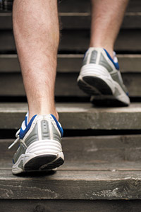 Heel strike places the foot in a supinated and semi-rigid position, shifting the majority of contact force to the lateral foot. In walking, supination normally reaches 2 degrees and up to 4 degrees in running. The continuation of this process moves the foot structures from a high-arched through a more neutral position, against the resistance of the maximally rigid, temporarily supinated foot. Flattening the arch dissipates energy and provides major shock absorption with each step.
Pronation begins as body weight transfers over the foot. The arch flattens to dissipate shock while the midfoot joints "unlock," allowing the foot to adapt to any uneven terrain. Ideally, the amount of healthy pronation should be limited to 8 degrees while walking and up to 12 degrees while running.1
Midstance prepares the foot for dynamic propulsion at toe-off. The foot must recover from pronation by supinating, relocking the mid-tarsals and shifting weight back toward the lateral foot. The foot must instantaneously transform from a fluid adapter to a rigid lever. Since both functions are equally important, this highlights why there is not one ideal or "neutral" position that could accommodate the variable demands placed on the feet.
Tibial rotational movements are considerable for a hinge joint. At heel strike, the tibia is externally rotated. During foot pronation, the tibia internally rotates and then returns to a position of external rotation up to and during toe-off. Limiting excessive rotational strain on the tibia is important for reducing or preventing many common conditions involving the knee.
Spinal reaction to movements in the feet. In a normal resting stance, the spine balances on a level sacral base created by legs of equal length and level femoral heads. During normal gait, steady, level motion is made possible by significant lateral displacements of the center of mass found near the pelvic bowl. This shift to the weight-bearing side helps maintain body weight centered over the hip.
Meanwhile, the weight-bearing ilium rotates anteriorly. Asymmetrical hyperpronation or any other cause of leg-length inequality increases time spent in anterior pelvic rotation. Consequently, lumbar lordosis is increased, putting excessive pressure on the posterior spinal joints.
The patient is not only likely to experience increased facet irritation, but also to compensate with an increase in the thoracic kyphosis and with anterior weight-bearing of the cervical spine. In fact, Cailliet says that for an inch of abnormal anterior cervical translation, there is a corresponding 10-fold increase in cervical spine muscle effort.2 This postural presentation of the classic chiropractic patient sets the stage for injury, fatigue and degenerative arthritis.
Interference and Dysfunction
Such far-reaching, complex motions of the gait cycle indicate the extent of stress that can result from interference with the timing, sequence and necessary rotation of movement. A muscular link between low back pain and the subsequent development of cervical symptoms supports this "chain theory" of reactions throughout the musculoskeletal system.3
Since muscle dysfunction can either be a direct source of pain or complicit in pain referral, there is an emphasis on balancing activity among groups of muscles for the prevention and management of pain syndromes.4 You will often note a strength difference between agonists and antagonists. Generally, trunk extensors are stronger than flexors.5 Furthermore, it is understood that some muscles are prone to weakness and others to tightening when injured.
Improperly functioning muscles also affect normal transmission of proprioceptive feedback, another mechanism in the kinetic-chain relationship.6 Chiropractic adjustments of the spine improve proprioceptive input by normalizing joint alignment and muscle tonus. Furthermore, because the feet contain approximately one-quarter of all the body's joints and therefore, a concentration of proprioceptive fibers, it becomes logical to conclude that support of the postural foundation using stabilizing orthotics will enhance balance and muscle coordination.7
Addressing Distortions Through Stabilization
It's advantageous and efficient to keep the center of gravity balanced near the midline and center of the pelvis. Therefore, any distortions in the lower extremity must automatically result in an equal but opposite distortion pattern above the pelvis. In fact, it may not always be easy to tell which distortion started first, but it is more important to realize that without correcting both, any correction will only be temporary.
Once you have stabilized the pedal foundation with orthotics and adjusted the associated distortion patterns, it is essential to retrain the muscular system to help maintain your corrections. For best results, simply have the patient perform an appropriate exercise in the position of function (position in which the distortion pattern was noted).
When patients don't respond to your chiropractic care as expected, look for a collapse in the arches of the feet or for habitual postural distortions. Unless treatments are based on a global, top-to-bottom analysis, results will likely be short lived. Successful treatment will combine specific adjustments to counteract the patterns of joint dysfunction, combined with rehabilitation of specific muscle groups and support for deficient structures, including the feet and lower extremities.
References
- Kapandji IA. The Physiology of Joints, Volume 2. New York, Churchill Livingstone, 1970.
- Cailliet R. Neck and Arm Pain. Philadelphia: F.A. Davis, 1981.
- Horal J. The clinical appearance of low back disorders in the city of Gothenburg, Sweden. Acta Orthop Scand, 1969;118(suppl):15.
- Lewit K. Manipulative Therapy in Rehabilitation of the Motor System. London: Butterworth, 1985.
- Langrana NA, Lee CK, Alexander H, Mayott CW. Quantitative assessment of back strength using isokinetic testing. Spine, 1984;9:287.
- Jull GA, Janda V. Muscles and Motor Control in Low Back Pain: Assessment and Management. In: Physical Therapy of the Low Back. New York: Churchill Livingstone, 1987.
- Stude DE, Brink DK. Effects of nine holes of simulated golf and orthotic intervention on balance and proprioception in experienced golfers. J Manip Physiol Ther, 1997;20:590-601.
Click here for previous articles by Mark Charrette, DC.