


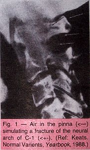
There are several occasions when there appears to be a fracture of the posterior arch of the atlas. It is important to identify whether or not a fracture is real or projectional to avoid unnecessary procedures and stress to a patient.
There is also a developmental anomaly that may give the appearance of a fracture of the posterior arch. This entity is absence of a portion of the neural arch of C1 on one side. If oblique views are taken, one side will appear normal and intact; the other side will demonstrate a cleft in the arch area, the region where the posterior arch connects with the body of C1 (Figure 2).
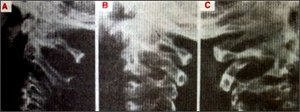
Fig. 2 - Unilateral absence of a portion of the neural arch of C-1. A, defect seen in a lateral protection. B, defect seen in oblique projection. C, normal side for comparison. (Ref: Karasick S, Karasick D, Wechsler RJ: Unilateral spondylolysis of the cervial spine. Skeletal Radiol 1983; 9:259.)
The other finding where there appears to be a fracture is due to faulty positioning of the head (a slight tilt of the head to one side). This may cause the posterior arch of C1 to appear to have bilateral defects in the arch (Figure 3).
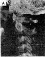
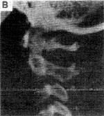
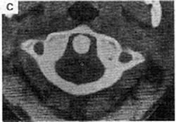
Fig. 3 - An appearance similar to that in the preceding figure may be produced by faulty positioning. A, apparent defects in neural arch of C-2 in off-lateral projection. B, defects now seen in true lateral projection. C, CT shows the neural arch to be intact. (Ref: Keats: Normal Varients, 4th Edition, Yearbook, 1988)
Many of these findings can be avoided if close attention is made to positioning the patient in the lateral projection. However, at times, this may be difficult to achieve, due to the patient's muscular spasms and true anomalies. Oblique views can be very helpful to rule out true fractures. If there is still a question, refer to a radiologist. Often a radiologist can give the final answer without having to perform more expensive studies, such as a CT scan or bone scan.
Deborah Pate, DC, DACBR
San Diego, California
Click here for more information about Deborah Pate, DC, DACBR.