The "tracer kinetic assay" method utilizes a radiolabeled, biologically active compound (tracer), and a mathematical model that describes the kinetics of the tracer as it participates in a biological process similar to, but far more sophisticated than, the radioisotopes in bone scan. The tissue tracer concentration measurement required by the tracer kinetic model is provided by the PET scanner, with the final result being an image of the anatomic distribution of the biological process under study.
Radiolabeled tracers and the tracer kinetic method are employed throughout the biological sciences to measure numerous biologic processes such as blood flow; membrane transport; metabolism; drug interactions with chemical systems; and marker assays using recombinant DNA techniques. Again, I point out that though most of the process is beyond my comprehension, this imaging tool is available and utilized regularly to evaluate patients for ailments such as neoplasms, cardiac viability and brain disorders.
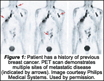
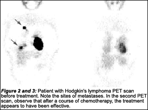
The radioisotopes used in PET are 11C, 13N, 15O and 18F - the radioactive forms of natural elements. These radioisotopes pass through the body, emit radiation and collect in various organs targeted for examination. A computer reassembles the signals into actual images. The PET scan can be used to determine if a neoplasm is malignant, and if there are other areas of metastases. It also can evaluate whether the treatment has been effective. It is regularly used in the evaluation of lung, colorectal, breast and prostate cancers.
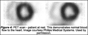
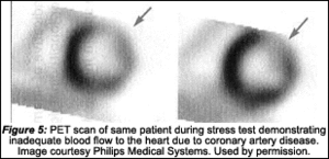
PET is the most accurate test to determine the viability of heart tissue for revascularization. It is presently the best method for determining the presence of Parkinson's disease, and is used to differentiate Alzheimer's from other types of dementia or depression. It also can effectively demonstrate the source of many common cancers, and heart and neurological diseases. PET is a proven diagnostic imaging modality that displays biological function in organ systems in the human body in a way never before available.
The following images illustrate how PET is used to diagnose and manage disease processes:
PET is a powerful diagnostic imaging modality that demonstrates the metabolic function in the organ systems, doing so within a single image. It often replaces multiple procedures and allows for monitoring the body's response to treatment. This modality is still developing, so we do not know yet what all its possibilities are.
As clinicians, we need to keep informed. Technology does not stand still.
Deborah Pate, DC, DACBR
San Diego, California
Click here for more information about Deborah Pate, DC, DACBR.


