My point is that most of us, including practitioners and patients, usually wait until we're hurting before doing anything about it.
I was taking my mother to her chiropractor for treatment (I long ago gave up treating relatives, especially parents - friends are okay).
"I don't understand why I'm having so much pain in my back," my mother said to me. This was after both my colleague (the treating chiropractor) and I had discussed her degenerative disc and joint disease just two days before, and on numerous occasions over the years!
She has two chiropractors she receives treatment from when she decides she is hurting. She won't go in for care when she isn't symptomatic. She thinks it's not worth her time. My mother is unable to drive because she doesn't see more that two feet in front of her, if that, but she does have access to transportation at any time. We go when she feels she needs to go. I suspect that you can relate to this scenario from your own patients and family.
Instead of getting frustrated, I decide to go the extra mile and explain once more. I find some visual material to try to explain what I think is her problem. There are many chiropractic products out there that one can use, but they require the ability of your patients to see well enough to read. I thought I would share with you some of the explanation that I gave her. Maybe some of these images will help some of your elderly patients understand better what may be aggravating their conditions.
The images in this article are from some of the work I did over 15 years ago in a fellowship program in osteoradiology. One of the research tools we used was to correlate the radiographic findings with the gross pathology. We used cadaver specimens that had known pathology (or even just degenerative disease), and imaged the specimen with x-rays and CT or MR. Then the region of interest was sliced with a band saw and evaluated by a pathologist. The radiographic images were exactly the same as the gross pathologic slices, with some minor changes due to freezing of the tissue. (To saw through the tissues, they needed to be frozen.)
I had all these old prints I had produced, and thought that they might help explain the degenerative process to my mother. (By the way, she has seen quite a lot of the chiropractic patient education literature because she has taken many courses provided by the Florida Chiropractic Association to train people as chiropractic assistants. Why do you think she did this? I'll give you one guess.) I know that this patient has had more than one report of findings regarding degenerative disease. This is how I explained it to my mother, luckily someone else's patient.
Instead of describing the findings on the patient's films and pointing out the problems, I decided to let the patient tell me what was wrong by first showing her what a normal spine looked like; and how the vertebral bodies and facets performed as a motor unit to provide the support and mobility that allows us perform the activities of daily living that we just assume we take for granted.
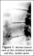
First I showed her a print of a normal lumbar spine:
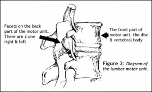
I explain the motor unit in very simple terms. For example, I explain that there is a disc with two left and right joints that make up the motor unit. The vertebral body and disc are the front half of the motor unit and the two vertebral joints are the back half. These two parts are its main structures. (You can fill in the gaps if you'd like, but don't get too complicated!)
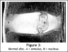
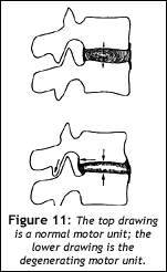
I show her the two facets, one right, one left, on the back of the motor unit, and the disc vertebral body on the front of the motor unit. Next, I show her a normal disc and explain that the disc is something like a sponge or, even better, a tire that absorbs and cushions the vertebral bodies and joints so they don't jam together and grate bone on bone. I explain that the fluid in the disc acts like a sponge that absorbs stress.
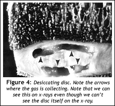
Then I showed her a desiccating disc.
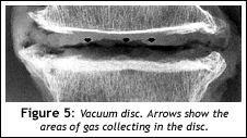
In Figure 4, look at the dark areas in the disc; those are areas of gas in the disc where there should be disc and fluid. Remember that the disc uses fluid pressure to help it bear the stress and keep the vertebral bodies and joints apart. Those areas you see with the gas are signs that the disc is not absorbing fluid properly.
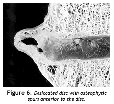
Next, I show my patient a really dried, crumbled-up (desiccated) disc and asked her to tell me if she understands the relationship between the sponge and the tire and the disc used in my earlier example. I also show her an old sponge that was pretty dried-up. She did not like the looks of the truly desiccated disc or the yucky sponge.
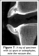
In Figure 6 note the spurs anterior to the disc. My mother notices these structures right off and wants to know what that was even before looking at the desiccated disc. These are bony formations that the body creates in an attempt to stabilize the ever-flattening and desiccating disc. This we can also see on plain x-rays, even though we can't see the disc itself.
The patient is impressed that the disc material extends out as far as the spurs do on the x-ray; she takes to the idea of the disc being the sponge; or that the flat tire or compressed sponge is spreading out around the vertebral body like a pancake; and a light seems to go on! This is not a new concept or idea, but I think some people just don't seem to correlate the two. (For whatever reason, I think I might have gotten through this time.)
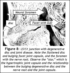
I next show her a specimen of a L/S junction with the desiccated disc and marked degenerative facets.(see Fig. 8)
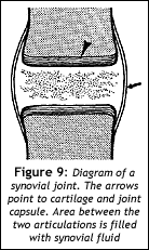
I think that once the patient has the concept of the tire or sponge, one can go to the facet joint and explain the degenerating process using those concepts. The facet joints are similar to all the other synovial articulations in the body. They are paired and allow flexion, extension and rotation; even in the lumbar spine there is some allowance for rotation. From that point, I explain the synovial joint, and show the patient a diagram of the joint.
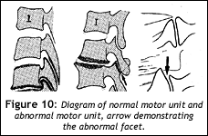
I start to hurry up with my explanations because I can see she was getting bored of the anatomy lesson. She wants her symptoms to just go away and go along with her normal activities of daily living without any discomfort. (Don't we all?) I show her a diagram of part of the lumbar spine with normal and abnormal motor units; changes on the abnormal one are consistent with degenerative disc.
I tell mother that the facet joints also lose their joint space by the wearing down of the cartilage forcing the joint capsule, which keeps the synovial fluid and the joint intact to work harder at keeping the unit together. In fact, the joint capsule sometimes works so hard at taking on the extra stress of keeping this joint together that the body starts to form osteophytes, or spurs around the joint capsule in an attempt to hold the joint in its normal position. Of course, when this starts to happen, the joint begins to lose its ability to move normally and again, the whole motor unit starts to break down.
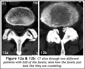
I probably should have ended my discussion here, but I didn't. I still had some of my mother's attention and wanted to complete the motor unit concept, i.e., that the disc and facets are dependent upon each other to balance the biomechanical stress of normal motion in the spine. To complete the facet idea, I showed her a CT scan of two different patients with degenerative joint disease in the lumbar region. Unfortunately, the CT slices are in the axial plane and the x-rays are in the sagittal plane, and it is confusing for most lay people to tell one plane to the other. I used the CT for the facets, because the lateral lumbar view does not demonstrate the joint spaces in the facets very well, and I thought I might be opening a can of worms to show a person the oblique view of the lumbar spine with a "Scotty-dog" configuration. I'm not certain which is better; you might want to experiment with some patients.
Since my mother has some difficulty with the different planes, I decide to just show her her own x-rays to see if she gleans any information from this discussion. I ask her to identify what might be degenerating in her spine. Amazingly, radiology is so easy; once you know what normal looks like anyone can see abnormal changes. This, and naming the abnormality (depending on who saw it or more likely published their findings first) is all that is required. The radiologists that follow are stuck with having to learn what the first person called the condition.
The patient will begin to understand what is happening and also may comprehend that this is a condition that is not going away. Once one motor unit starts undergoing this process it will affect the adjacent motor units; as the process progresses, it involves more motor units, and so on. This progression may be slow, but over time it will very likely cause symptoms. There are many ways to interrupt this progression. Some ways may work better than others, and in some situations, the degeneration may be so severe that only surgical intervention will alleviate the symptoms for some time. But biomechanically, even if just one motor unit isn't moving properly, eventually there will be a change in the distribution of the biomechanical stresses and some part of a motor unit will be overstressed, and the degenerative process will begin.
I think it is important to know when to stop talking and allow the patient to digest the information. If the patient wants more information, you can oblige. Many people are not truly interested in taking responsibility for their own health; this should be sufficient to maybe give them some understanding of what is happening to them. I think that if the population were to understand these simple concepts and (which is a big "and") were intent on managing their own health, there would never be enough chiropractors to fill its demand.
By the way, my mother repeated that she could not understand why she still had back pain. I showed her the old beat-up sponge.
"Yes, I know all about that disc-and-facet-and-motor-unit stuff," she said, but I still would like to know how I happened to get DJD since I'm only 79."
Deborah Pate,DC,DACBR
San Diego, California
Click here for more information about Deborah Pate, DC, DACBR.


