The most common level for a spondylolysis is L5.
The best view to determine the presence of a spondylolysis in on the oblique view. If there is a question as to whether or not there is one present, a CT scan will demonstrate this region very clearly. If a possible recent fracture is suspected a bone will demonstrate whether or not the spondylolysis is recent.
Spondylolisthesis is defined as a subluxation of one vertebral body on another; displacement generally is understood that the superior vertebral body is displaced anterior in relationship to the inferior vertebral body.
Spondylolisthesis may be classified as isthmic, degenerative, dysplastic, traumatic or pathologic. In isthmic spondylolisthesis, the cause of the spondylolisthesis is due to a spondylolysis of the pars, and therefore allows the anterior slippage of one vertebrae on another. Dysplastic spondylolisthesis is often due to developmental hypoplasia of the pars, which may be elongated, allowing for the anterior slippage of one vertebrae on another. Traumatic spondylolisthesis is almost always due to a fracture of the posterior arch which allows for slippage of the fractured vertebrae on the adjacent segment. The best example is the "hangman's fracture." Degenerative spondylolisthesis is due to alteration in the biomechanics of the facets, which generally demonstrated marked degenerative changes and allow for the spillage of one vertebrae on another. Neoplastic processes can cause destruction of a vertebral body, again allowing for the displacement of one vertebrae on another, which is termed a pathologic spondylolisthesis.
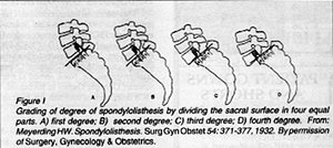
It is important to note what type of spondylolisthesis is present for clinical management of the patient. There is also a grading system for determining the stability of a spondylolisthesis. This system I am sure everyone is aware of, however, it is more important to determine if there is in fact a progression of the anterior slippage. Any progression of the anterior slippage is even more important than the grade of spondyloslisthesis. If there is radiographic evidence of an increase in the anterior displacement, that alone is enough to document instability.

Grading of degree of spondylolisthesis by dividing the sacral surface in four equal parts. A) first degree; B) second degree; C) third degree; D) fourth degree. From: Meyerding HW. Spondylolisthesis. Surg Gyn Obstet 54: 371-377, 1932. By permission of Surgery, Gynecology & Obstetrics.
Deborah Pate, DC, DACBR
San Diego, California
Click here for more information about Deborah Pate, DC, DACBR.


