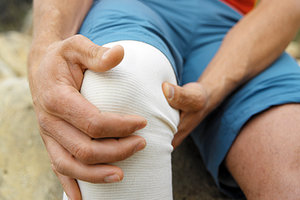
We have all seen patients with medial knee pain that either has no traumatic origin or lasts well beyond when it should be resolved. How can we help these patients? Here is an overview of clinical scenarios and how we can provide conservative care.
First, you need to rule out ligament or cartilage pathology. If the patient has a complete tear of the anterior cruciate ligament (ACL) or the medial collateral ligament (MCL), they are less likely to respond to conservative care. Imaging, even advanced imaging such as MRI, is still a gray zone; still not perfect.
Do they have a meniscal tear? A newer and more sensitive physical exam test is the Thessally test.1 Can they stand on the involved leg and twist back and forth without pain? They are dynamically challenging the meniscus through weight-bearing rotary stress.
Lots of patients have some degree of DJD of the knee. That is not necessarily the cause of their pain. Our job is to search for and correct the fixable lesions.
The most common muscular or stability contributor to medial knee pain is a lack of control of medial motion in the knee, creating valgus stress. Test this via the lunge, or stepping down from or up onto a small stool. If the knee fails into valgus, the key therapy is training the hip abductors. Use the clam shell, functional clam shell and/or a crab walk with band resistance. If you are not testing for and using this simple rehab, in my opinion, you are not up to a minimal standard of care.
 The valgus knee is a huge contributor to not only medial and anterior knee pain, but also to an increased risk of ACL tears. Despite all the advances in surgery, once the ACL is torn, the patient is very likely to have accelerated degeneration of the knee. Prevention is key.1
Beyond the Basics: Thinking Outside the Box
If you are following an assess, treat, reassess model, you need to start with an indicator. I often look for a tender point, which may be over the medial joint line or over the pes anserine. A functional indicator would be the ability to do a lunge without either pain or valgus movement of the knee.
Once you have started with rehab and/or pain-relieving modalities and the patient is not responding as quickly as you hoped, what is next? Don't just keep doing the same thing. Pain is a liar; he who treats the site of pain is lost. Do you aspire to excel? Do you like the challenging cases? Do you want to be the go-to doctor for the patients who are not getting well?
Let's discuss 11 types of potential knee dysfunctions, including assessment and treatment. These 11 patterns are amenable to manual care and are listed in proximal-to-distal order for simplicity of examination. (I want to credit a few of my many creative teachers here, including Craig Liebenson, DC, Tom Hyde, DC, Paul Chauffour, DO, Jean Pierre Barral, DO, Warren Hammer, DC, Luigi Stecco, PT, and Lucy Whyte Ferguson, DC.)
11 Factors That Can Contribute to Medial Knee Pain
- Check the fascia of the kidney and correct if it is stuck.
- Check and correct the sacroiliac and especially the anterior part of the pelvic ring, the pubic symphysis.
- Check hip joint motion.
- Assess, treat and rehab the adductors; you'll often find a combination of weakness and tightness.
- Check the popliteus; it is a small but significant stabilizer of the knee.
- Check for joint restriction within the knee joint complex.
- Check the medial ligaments and tendons of the knee.
- Check both the medial fascial lines and the lateral fascial lines of the lower extremity on both the involved and non-involved leg.
- Check the saphenous nerve as an irritated peripheral sensory nerve.
- Check the tibia itself for intraosseous restriction.
- Check the foot for potential issues.
In reviewing this list, note that almost all of these factors are connected with the medial fascial lines. The medial knee is midline and most of the components that affect it are midline as well. I think of both joints and fascia as contributors to fascial dysfunction. If this is a new idea to you, look at Tom Meyers' anatomy trains and/or Stecco's fascial manipulation; both systems have lines or sequences of fascial points to assess.
When I am doing this kind of outside-the-box work, I like to quickly recheck the tenderness indicator after each correction. Which piece was significant? There is no perfect system; it is often a combination of factors that creates pain the body can't resolve.
1. The Fascia of the Kidney
Check the mobility of the kidney, and correct if it is stuck. I wish more DCs were familiar with visceral manipulation.2 The kidney is another near-midline medial structure with wide-ranging effects. This is not kidney toxicity or kidney pathology. This is not dehydration. This is a fascial problem, with the kidney usually dropped inferior.
One of the few peer-reviewed articles on kidney manipulation focuses on its effects on the lower back. If lower back pain is a little known effect, restriction of the kidney affecting the knee is even less known. I have had many knee patients in whom this was a key factor.
2. Sacroiliac & Pubic Symphysis
Our focus here is on the pubic area. Start with visual inspection. Place your two thumbs side by side on the top of the left and right pubic symphysis. If in lesion, you'll observe that one side is superior or inferior. The second major indicator is tenderness, either at the top or the bottom of the medial pubic symphysis.
Keep in mind that you are touching a sensitive, sexually charged area. Use the appropriate statements. Explain what you are examining and why, and ask specific permission to touch the pubic area.
I will not detail correction here. I will say, don't thrust, and don't go for an audible release. Muscle energy and counterstrain have elegant techniques for this area. A simple push-pull move sometimes works, and you also need more specific techniques.
3. The Hip
I have written extensively on this.3 Start by checking hip range of motion. I prefer supine at 90 degrees of flexion; others test it prone. I always check one side against the other, as normal internal rotation varies immensely. What happens when the hip cannot internally rotate? The hip is stuck in external rotation and every step overloads the medial knee. A clinically significant hip that lacks internal rotation will also display tenderness over the head of the femur and weakness of the hip flexors, as tested above 90 degrees.
I love Lucy Whyte Ferguson's correction. It works by eccentrically activating the adductors, so you get a three-for-one benefit. You are correcting hip motion, stretching the adductors and activating the adductors. Big win.
4. The Adductors
The adductors fit into the "most missed" category. So many of us are afraid to touch that deep into the groin. I tend to find trigger points or tender points at various levels, top to bottom, in the adductor region.
The adductors originate from the pubic symphysis region. If the adductors are off, especially at the upper end, I will also check the obturator foramen, the deep insertions of the sacrotuberous ligaments along the ischium, and the alignment of the pubic bones.
Don't just think tight, think weak and tight. Isometric activation of the adductors can help wake them up. You can modify a lunge, as foundation training's "woodpecker" exercise has done, to focus on isometric or eccentric adductor activation.
5. The Popliteus
The popliteus is an oblique sling behind the knee. Think of it in posterior as well as medial knee pain. Patients who have difficulty kneeling, squatting, running hills or going down stairs should be evaluated for popliteal tendon disorders. (Thanks to Dr. Tom Hyde for teaching me the significance of this muscle.)
You have to be accurate with your palpation. Check both the origin and the insertion areas. Motion or positioning will help make the dysfunction more obvious. The muscle is less palpable with the knee at 90 degrees of flexion; more obvious at about 15 degrees of flexion, as you begin to put a stretch on the popliteus. The origin is palpated just anterior and medial to the lateral hamstring tendon, on the posterior distal lateral femur. The insertion is behind the medial knee, just lateral to the medial gastrocnemius tendon.
For both the origin and insertion, don't just push P to A. Hook your finger and pull or push toward the attachment for a better feel. When involved, it will be tender. Treat with instrument-assisted soft-tissue mobilization or other fascial work. Dynamic treatment during motion, as used by FAKTR, can help make your soft-tissue work more effective.
6. The Knee Joints
Yes, we do sometimes find the need for knee joint manipulation. Even though it is a lateral structure, the fibular head can be significant for medial pain. Use motion palpation. Does the fibular head resist anterior or posterior motion?
I like to use instrument-assisted adjusting here as a simple correction. I always fine tune the correction to go slightly medially or laterally with my thrust. The other significant knee joint fixation is a rotary fixation of the tibia. Always compare left to right. I like a simple, quick thrust in the direction of resistance. It does not take a lot of force and you will rarely get an audible release.
7. Intraosseous Tibia Restriction
This is another factor too few of us are trained in.4 Any long bone is designed to bend. If it cannot bend normally, it probably cannot absorb shock appropriately.
I won't try to describe this assessment and correction in two sentences. I wrote two articles many years ago on this topic. As I re-read them, I am glad to see that they have stood the test of time and do a good job of describing both assessment and correction.4-5 I also recommend you read an interesting article about an osteopath using thermal imaging to "see" these intraosseous strains.6
8. The Medial (and Lateral) Lower Extremity Fascia
I am talking about the whole of the lower extremity, following the medial and lateral lines from the pelvis down to the foot. Stecco has elegant maps of where the most likely fascial densities are likely to be found. If you use Travell's model, check those classic trigger points. You should also check the medial plantar fascia of the foot, and the psoas and iliacus, to be complete.
Release any tender points with your favorite fascial release work. Stecco emphasizes establishing a balance between medial and lateral, and left and right.
9. The Medial Collateral Ligaments and Tendons That Cross the Medial Joint Line
Maybe you addressed this already when treating the medial fascia. I mentioned that complete ligament tears are unlikely to respond to our care. Here, I am thinking of overstretched, clinically lax ligaments. Start with your IASTM as a pro-inflammatory soft-tissue technique, attempting to get the body to lay down new collagen tissue, and see if the patient responds.
The fitter person, willing to work out and build muscles and ligaments, is often a better candidate for this. If the ligaments are not responding, or if the results are temporary or inadequate, this patient may be a candidate for prolotherapy injections.
10. The Saphenous Nerve
My most recent article was on pain from sensory peripheral nerves.7 If the saphenous nerve, at its infrapatellar or medial crural cutaneous branches, is irritated and swollen, it can cause medial lower leg pain, which may be experienced as medial knee pain or as a neuropathic leg pain.
Try laser or LED light therapy, dextrose cream, e-stim, tissue approximation or counterstrain. These nerve irritations are less likely to respond to deep tissue. Again, if the results are temporary, the patient may be a candidate for peripheral nerve injection therapy, also known as neuroprolotherapy.
11. The Foot
If the patient pronates, orthotics may help protect their medial knee. When you have medial knee pain, the first place to check in the foot is the medial side. What happens there? The big toe tends to hurt and DJD often develops at the first MTP joint.
The joint restriction that commonly contributes to this is more proximal, at the first and second metatarsal-cuneiform joints. Typically, this area is stuck inferior and needs release upward. If the subluxation continually recurs, think of orthotics; think of how to accommodate the foot with hallux rigidus. Check the rest of the foot as well, especially around the talus.
There you have it: 11 different things to check when evaluating / treating medial knee pain, beyond the basics of ruling out pathology and basic rehab. I'll bet a few of these are either new to you or at minimum, not part of your exam routine. You will never be bored if you practice this way.
References
- The Thesally test (video demo including description and references).
- Heller M. "Visceral Manipulation." Dynamic Chiropractic, April 15, 2013.
- Heller M. "The Hip: Myofascial and Joint Patterns." Dynamic Chiropractic, May 7, 2007.
- Heller M. "Intraosseous Restrictions." Dynamic Chiropractic, Nov. 5, 2001.
- Heller M. "The Tibia and Femur: Long-Bone Intraosseous Restrictions." Dynamic Chiropractic, April 9, 2005.
- Muntinga E. "Detection of Intraosseous Strains in the Adult Male Tibial Bone: Osteopathic Palpation and Thermography." Thesis protocol, Oct. 23. 2011.
- Heller M. "Assessing and Treating Peripheral Sensory Nerves." Dynamic Chiropractic, June 1, 2014.
Click here for more information about Marc Heller, DC.


