A major soft-tissue problem with a variety of unproven treatments is patellofemoral pain syndrome (PFPS). PFPS is a general term that refers to patellofemoral dysfunction due to knee extensor overuse lesions and mechanical disorders causing articular pain (arthrosis), leading to degenerative joint disease or malalignment.
In a recent study by Tyler, et al.,2 excellent results occurred with PFPS if the patient had a combination of weak hip flexors, and tight iliotibial band (Ober test) and iliopsoas (Thomas test). Patients with this combination of dysfunction and PFPS were successfully treated 93 percent of the time. The patients in this study experienced anterior or retropatellar knee pain from at least two of the following activities: prolonged sitting, stair climbing, squatting, running, kneeling and hopping/jumping, or persistent anterior knee pain for at least four weeks with no apparent reason. Patients with intra-articular problems, ligament involvement and even tenderness over the patellar tendon, iliotibial band or pes anserinus tendons, hip- or lumbar-referred pain, previous knee surgery, or degenerative joint disease were excluded from this study.
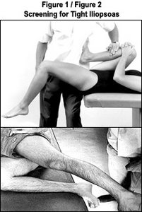
The iliopsoas was tested with a modified Thomas test position (Fig. 1). For the test, the patient sits at the edge of the table, with the contralateral limb held in the knee-to-chest position and his or her back against the table. If the thigh is horizontal with a rigid end-feel or above the horizontal, the iliopsoas (hip flexor) is considered tight.
The Ober test measures the tightness of the iliotibial band, or ITB (Fig. 2). The patient lies on the uninvolved side with the knee flexed. The upper leg can be flexed 90 degrees or extended. It is important for the examiner to stabilize the hip at a right angle to the table as he or she grasps the upper thigh/leg, abducts it and allows it to lower. If the extremity remains horizontal and rigid or above the horizontal, the iliotibial band is considered tight.
 Hip-flexion strength can be measured with the patient in the sitting position, then being asked to raise the thigh eight inches above the table and to maintain the position. The examiner can press down or use a hand-held dynamometer as he or she forces the thigh toward the table. Three tests should be given for both thighs to measure for strength.
Positive Ober and Thomas tests have been related to an anterior pelvic tilt, which may create femoral internal rotation. Femoral internal rotation influences patellar alignment and kinematics.1 "Increasing the flexibility of the hip flexors and ITB would allow the pelvis to rotate posteriorly, creating relative femoral external rotation and helping to align the patella in the trochlear groove of the femur."1,2
By increasing the strength of the hip flexors, a more stable pelvis will occur during gait, allowing it to act eccentrically to prevent the pelvis from going into an anterior pelvic tilt and concomitant femoral internal rotation. Strengthening the iliopsoas, an external rotator of the femur, will help align the trochlear groove with the patella. Increased flexibility of the hip flexors and ITB can reduce the tension of the lateral retinaculum and allow the patella to track normally. Lateral patellar tilt and displacement can be due to femoral internal rotation, not patellar motion.2,3
There are many ways of increasing the flexibility of muscles, such as postfacilitation stretch, postisometric relaxation, myofascial release, active release, Graston technique, neuromuscular re-education, and numerous stretching methods.
References
- Powers CM. The influence of altered lower-extremity kinematics on patellofemoral joint dysfunction: a theoretical perspective. J Orthop Sports Phys Ther 2003;(33)11:639-646.
- Tyler TF, Nicholas SJ, Mulaney MJ, McHugh MP. The role of hip muscle function in the treatment of patellofemoral pain syndrome. Amer J Sports Med 2006; (34)4:630-636.
- Lee TQ, Morris G, Ssintalan RP. The influence of tibial and femoral rotation on patellofemoral contact area and pressure. J Orthop Sports Phys Ther 2003;33:686-693.
Click here for previous articles by Warren Hammer, MS, DC, DABCO.


