


Most of the time, a snapping sensation in the hip is painless and, except for the noise or even palpating it, the lack of symptoms results in the opinion that it does not require treatment.
The condition has been classified into three categories: external, internal and intra-articular.2 External causes are the posterior iliotibial band, the anterior gluteus maximus and trochanteric bursitis. Internal causes are the iliopsoas tendon, iliofemoral ligament and hamstrings. Intra-articular causes are labral or ligamentum tears, loose bodies, synovial chondromatosis, displaced fractures and capsular instability.3 Many patients can voluntarily demonstrate the snap.
The most common and easiest type of hip snapping to diagnose is external, which involves the posterior iliotibial band (ITB) or the anterior aspect of the gluteus maximus as it travels over the greater trochanter during hip flexion and extension. The thickening of the posterior ITB and anterior fibers of the gluteus maximus causes catching and pain.3 This external type is found most often in cyclists and runners.
A functional test for the ITB is to have the patient lie on their side and move the lower extremity through extension and flexion of the hip, and then through internal and external rotation. Listen or palpate for a snap and inquire whether this causes pain (although a snap does not necessarily have to occur). This test also can be done passively with the patient lying on the noninvolved side, by extending and abducting the hip with a straight leg, producing a snap over the greater trochanter. If the lower extremity remains abducted when the patient is told to relax, this is evidence of a tight ITB, similar to an Ober test. An Ober test should reveal a tight ITB.4
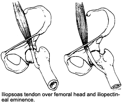 The most common internal type of snapping hip is due to the iliopsoas muscle. To test for snapping, start with the FABER position and then passively move the hip into an extended, adducted and internal rotation. The figure below depicts the iliopsoas tendon sliding over the femoral head and iliopectineal eminence from lateral to medial, with movement from flexion, external rotation and abduction (left) to a more extended, internally rotated and adducted position (right). This test puts tension load across the tensed iliopsoas tendon and also might involve snapping due to tension from the ilioinguinal ligament.2 Attempt to palpate the snapping over the anterior thigh during this test. Also test for a shortened iliopsoas.
The intra-articular type, due to the lesions stated above, may require diagnostic imaging, although there are functional tests that may lead you to the intra-articular causation (i.e., labral testing). Wahl, et al.,3 state that if there is labral pathology, there will be a "click," compared to the extra-articular "snapping," "vibration" or "locking." "Searing pain" or "grinding" may be due to mechanical incongruence caused by loose bodies, hip dysplasia or osteoarthritis.
Some ballet dancers have found that snapping their hip on purpose relieved their pain; thus, it could be that the stuck hip had to be snapped for relief.1 Some of the findings that might require treatment include leg-length difference (usually the long side is symptomatic), tightness in the iliotibial band on the involved side, weakness in hip abductors and external rotators, and poor lumbopelvic stability. Abnormal foot mechanics (e.g., overpronation) leading to increased femoral internal rotation should be evaluated.
An article by Konczak and Ames5 discussed a patient who was a runner with low back pain and anterior hip-popping. This patient achieved results by sideposture SIJ "diversified" manipulation and myofascial release to the psoas muscle twice weekly, for two weeks. The patient also was taught proprioceptive neuromuscular facilitation exercises of the psoas and iliotibial band muscles. He was instructed to substitute swimming instead of running on a daily basis.
References
- Winston P, Awan R, Cassidy DJ, Bleakney RK. Clinical examination and ultrasound of self-reported snapping hip syndrome in elite ballet dancers. Am J Sports Med, 2007;35(1):118-26.
- Allen WC, Cope R. Coxa saltans: the snapping hip revisited. J Am Acad Orthoped Surg, 1995;3(5):303-8.
- Wahl CJ, Warren RF, Adler RS, et al. Internal coxa saltans (snapping hip) as a result of overtraining: a report of 3 cases in professional athletes with a review of causes and the role of ultrasound in early diagnosis and management. Am J Sports Med, 2004;32(5):1302-9.
- Hammer WI. Functional Soft-Tissue Examination and Treatment by Manual Methods, 3rd edition. Massachusetts: Jones & Bartlett, 2007:259-309.
- Konczak CR, Ames R. Relief of internal snapping hip syndrome in a marathon runner after chiropractic treatment. J Manipulative Physiol Ther, 2005;28(1):e1-7.
Click here for previous articles by Warren Hammer, MS, DC, DABCO.