Probably nothing is more dispiriting to the life of a practice than to do harm while in the quest to do good. We are geared to be helpers and healers, and the acknowledgment that we are capable of making our patients worse is a sobering lesson.
A review of malpractice claims reveals that the single largest category of harm done is disc herniation. Other less common causes for harm are: failure to properly diagnose gross pathology (which often means failure to refer patients for further evaluation when indicated by primary complaint, history, physical findings or clinical course); poor-quality X-rays that are not of diagnostic value; fractured ribs; failure to adequately document physical findings, test results and patient progress; inadequate orthopedic/neurological testing prior to treatment; vertebral artery dissection leading to stroke; and disregard for poor treatment outcomes that should be considered red flags, necessitating re-evaluation prior to further treatment. A lack of informed consent to the possible dangers of spinal manipulation only exacerbates such cases.
 As regards disc herniation, many of us accept that acute spinal trauma or chronic spinal biomechanical impairment commonly accompanies some degree of disc compromise. Acute spinal injuries that tear or stretch paraspinal ligaments also may involve sufficient force to threaten the integrity of the disc's annular fibers, leading to disc weakness and bulge. Chronic spinal impairment often is occult in nature, with intermittent acute episodes. Such impairment of motion is thought to disrupt the normal cycle of daily disc dehydration/rehydration - a result of axial loading during the day and resorption of fluids at night - and results in the eventual desiccation of the disc. This "drying out" of the disc is the first sign of degenerative disc disease seen on an MRI.
It would seem, therefore, a prudent approach for practitioners of spinal manipulative therapy to assume that any biomechanical lesion (subluxation) he or she identifies through palpation also may contain a weakened disc. This would seem particularly so for patients over 35 years of age or anyone with a history of significant trauma, ponderous obesity or repetitive heavy lifting.
Many patients with disc herniation can identify the exact instant the prolapse occurs. These cases are more easily identified if there is an accompanying radicular neuritis with lost or diminished deep-tendon reflex, specific dermatomal dysesthesia (soft touch is first to go), and associated myotome weakness. Muscular atrophy, evidenced by disparate extremity circumference measurement, is a telling sign of significant protracted neural compression. Increased radicular pain with weight-bearing and forward-bent antalgic posture relieved by recumbence are other heralding characteristics of disc herniation.
Most of us have little problem identifying these cases in the clinic and often are rewarded by a confirming MRI. To try and manage these patients conservatively is an option, but outcome success with these patients is mixed. One encouraging development is that there have been reports of disc resorption on serial MRIs, and decompression traction has shown great promise in recent studies. Of course, should a practitioner attempt hands-on treatment, the patient should specifically be advised of the risk of spinal manipulation for increasing the size of disc protrusion. When I find frank neural deficit, especially with some degree of paralysis, I personally refer these patients for neurosurgical consultation. If they then choose to try conservative options, I always include some form of traction in the regimen of treatment.
While specific case histories are hard to come by, it is probably not these cases that are most involved in malpractice litigation. Rather, it is those cases with a more nuanced clinical presentation - less distinct neurological signs - that pose the greatest risk. Because of this, any peripheral manifestation, such as pain, numbness, tingling, burning or muscle weakness, should be approached with great caution. Quite simply, to treat these patients without having first performed a thorough neurological examination is to invite a malpractice suit. From my experience, while it is true that biomechanical impairment can create a peripheral neuritis, it should never be assumed as the only possible cause. Imagine a large posterior disc bulge with only a few outer ring fibers intact being taken to end-range-of-motion rotation or full P-A compression and then subjected to a sharp force of high velocity. Bingo! It happens all the time.
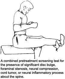
As we now realize, disc bulges are relatively common in the general population and often are completely asymptomatic. When they do manifest, they often mimic the pain of spinal joint segmental dysfunction. It is for this reason that I developed a screening test for all new patients that challenges the intracanal and nerve root structures for signs of "silent" inflammation or active compression. It has proven very effective in practice.
As you probably know, the sitting straight-leg-raise (SLR) test is superior to the supine (Laseque's) SLR test in that it adds the additional component of axial loading to the discs and vertebral structures during testing. Normal disc expansion with weight-bearing is only about 1 mm, but where there is a weakened outer ring, one would expect significant additional expansion. Thus, low back or leg pain with straight-leg raise may manifest only with the disc under axial compression.
Hard, forceful neck flexion, including the oblique planes, is a good test for disc protrusion or foraminal stenosis in the cervical spine. A positive sign would be anterior neck pain or the creation or increase of peripheral pain or dysesthesia. This maneuver has the additional effect of stretching taut the spinal cord, tractioning the nerve rootlets, spinal cord, and its attachments at the cauda equina. It also pulls the spinal cord up against the posterior wall of the vertebral bodies and discs.
The Valsalva maneuver, as we know, increases intracanal spinal pressure. It is helpful in screening for cord tumor. Edematous neural tissue located within the canal or adjacent to it would, therefore, also be compressed by the increased pressure, causing an exacerbation of existing pain signal or perhaps even bringing an occult neuritis to the surface. Do not, however, use this procedure with anyone suspected of having severe coronary disease, as it may illicit a heart attack.
It seemed to me that combining all three tests would be an excellent method to screen all new patients for the presence of hidden cord tumors, intracanal compression or paraspinal neuritis. It also has the advantage of helping to distinguish neural pain from capsular/facet pain. I begin the test with the sitting straight-leg raise, and then instruct the patient to hard neck flexion, finally adding the Valsalva maneuver - instructing the patient to inhale deeply and hold, followed by hard muscular contracture of the upper body. I may even then actively elevate the leg with one hand and press down on the flexed head with the other, to add additional stress to the combined maneuver. If there is an active neural compression within or adjacent to the spine, I want to know it.
With positive findings, I follow up with specific neurological testing and perhaps MRI exam. With a negative finding, I feel pretty confident I am dealing with a primary joint pain syndrome and proceed with my usual therapies.
In these days of demanded high-dollar malpractice coverage for participation in various health insurance schemes, and the reality of contingency fee plaintiff-lawyer arrangements, it only seems prudent to have a reliable, quick screening test for the presence of neural compromise or disc weakness. But, most importantly, it is our duty to our patients, first and foremost, to do no harm.
Dr. John Bomar, a 1978 graduate of Palmer College of Chiropractic, practices in Arkadelphia, Ark. He is a past board member of the Arkansas Chiropractic Association and a founding board member of the Arkansas Chiropractic Educational Society. Contact Dr. Bomar with questions and/or comments regarding this article via e-mail:
.


