Anterior Lower Cervical Subluxation Patterns
Most of us screen the lower cervicals from behind the patient as part of our palpatory exam, but I find that the anterior neck is rarely assessed or treated. We'll focus on the anterior restriction, where C6 or C7 resist posterior glide. You have to palpate sitting to find this problem. When the patient is supine, this lesion partially self-corrects and becomes impossible to feel.
I first started appreciating this area when I studied Dr. John Bandy's "cervical disc syndrome." Dr. Bandy uses weakness of the biceps to indicate C6 nerve root problems, the triceps to show C7 impingement, and the finger abductors to indicate C8 nerve problems. He assesses for a "stressed" position - usually flexion combined with lateral bending and rotation into the involved side - which further weakens these muscles. The muscles may test strong in neutral posture and only show weakness in the stressed position. When I started to use this protocol on my patients, subluxation patterns emerged.
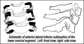
The subluxed vertebra is usually translated forward, lateral, and tipped inferior. In dynamic terms, it resists posterior glide, lateral to medial motion, and inferior to superior motion. This is a nonphysiological restriction, which does not follow the joint planes. This confused me for years, until I integrated Barral's understanding of this area. Barral talks about the anterior fascial pulls and their influence. When these fascial structures are tight and restricted, they pull the neck in an inferior direction. My next article (May 20 issue) will focus on the inferior fascial pulls and how to find and correct them.
| Related Literature Dr. Lewit speaks of the importance of key segments, mostly in transition zones, including the cervicothoracic junction, "where the most mobile section of the spinal column is joined to the relatively rigid thoracic spine." In Motion Palpation and Chiropractic Technic, Schafer and Faye state: "One of the two most common fixations in the lower cervical spine involves (restriction of) simultaneous flexion and A-P rotation." In The Thorax, Jean Pierre Barral emphasizes the fascial components pulling on the front of the neck. The chief complaint coming from lower cervical problems is often a referred pain felt in the upper thoracic region or experienced as shoulder/arm discomfort or sensory changes. Dwyer, Aprill, and Bogduk have done a beautiful job of outlining referral patterns from the cervical spine based on injection responses. Lower cervical dysfunction correlates with Janda's upper-crossed syndrome, in which the head carriage is forward in a "chin-poke" posture, the scalenes and SCM are hypertonic, and the deep neck flexors are inhibited. |
Eighty percent of these patients will have an inferior component to the lesion, resisting superior motion. Approximately 20 percent of the time the pattern will be forward, lateral, and superior - more along the lower cervical facet lines. It's quite significant that the usual pattern is nonphysiological - not along the joint planes. Since we don't have a facet line along which to adjust, we have to use low-force correction, and we must address the fascial restrictions.
Diagnosis and Palpation of the Subluxation
How do we find this problem? Screen the sitting patient's lower neck with gentle palpation using initial response testing (IRT), feeling for rigid areas over the front of the lower neck. In IRT we assess the very beginning of the motion rather than the end range. Go through the SCM muscle by pushing it gently backward to access the anterior part of the transverse processes. If you feel an arterial pulse, your contact is more medial than it should be. The cervical transverse process is a sensitive area on almost anyone, but it will be hypersensitive and rigid over an area of dysfunction. To motion palpate, passively flex the head forward and laterally bend it toward the involved side with one of your hands, while the palpating fingers or thumb of your active hand counter this motion by gently pushing anterior to posterior, and simultaneously pushing medially. You will feel a lack of movement at the significant level. Add your third dimension by determining whether the stuck vertebra resists superior motion, the common pattern, or resists inferior motion along the facet line. If the patient has a severely irritated cervical nerve root, lateral motion will not be tolerated, so just assess A-P and I-S motion in these cases.
Correction: Three Variations on Low-Force Adjusting
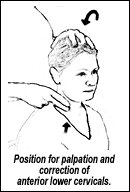
How do we adjust over the very sensitive anterior portion of the neck? I use these low force methods with the patient sitting. It's a lot like the palpation assessment. I'm either behind the patient, as pictured, or standing in front on the involved side. I begin by gently side-bending the patient's head to the involved side. I primarily use engage, listen, follow (ELF) to correct the restriction. I engage the elastic, soft part of the barrier with my thumb or index finger, then push gently and slowly posteromedially on the involved segment, while my upper hand simultaneously rocks the head anterolaterally toward the involved side. I fine tune for the usually superior direction of restriction. Once I have engaged the lesion, I listen for the exact 3D direction the tissues want to release toward, and follow to completion. This is a variant on direct myofascial release, gently and directly moving into the elastic barrier, pushing the C6 or C7 vertebrae in a posterior, medial, and superior direction. This is done slowly three to five times.
A second technique involves adding postisometric relaxation (muscle energy) to the same setup. I use this when my direct ELF doesn't seem to completely release the area. Begin as above and move directly into the barrier. When you reach the first barrier, have the patient very gently and isometrically bring the head laterally against your upper hand's resistance into opposite side-bending for four to seven seconds. This is done with minimal pressure. After the contraction, let the patient relax and follow with your direct mobilization toward the barrier. Repeat three to five times. One key is to take the segment just to the soft edge of the barrier. We do this by backing off slightly before you have the patient begin to contract. The other key is teaching your patients to contract their own muscles very gently. They always push too hard the first time. I coach them by saying, "Give me about one-fourth of that much pressure, and as soon as you feel my resistance, just hold, don't continue to try to push further."
A third technique, which can be combined with the other two, would be recoil. This is best described as engage and release. Engage the restriction in all three dimensions, pushing only to the soft part of the barrier, and then suddenly release your pressure. Unlike toggle- recoil, there is no thrust inward, just a sudden release away. This can be used at the beginning or end of ELF, or on its own for an acute case when motion cannot be tolerated here.
Always recheck after your correction. The area should be less tender, more mobile, and any muscle weakness should be gone.
Using these concepts may transform your approach to the cervical spine. Look to the anterior lower cervicals whenever the patient complains of pain in the lower neck, shoulder blade or upper back, or whenever an upper extremity pain or sensory change is not resolving fully.
My next two articles will address the fascial and muscular components of the anterior cervical syndrome.
Resources and References
- Greenman P. Principles of Manual Medicine, 2nd edition. Williams and Wilkins, 1996.
- Lewit K. Manipulative Therapy in Rehabilitation of the Locomotor System. Butterworth, Heinemann, 1991.
- Schafer RC, Faye LJ. Motion Palpation and Chiropractic Technic: Principles of Dynamic Chiropractic. Motion Palpation Institute, 1989.
- Advanced AK Seminars with John Bandy, 1987, Austin, Texas.
- Barral JP. The Thorax, Eastland Press, 1991.
- Heller M. Low-force adjusting techniques (including ELF). Dynamic Chiropractic, Sept. 1, 2001;19(18), pp. 32,34, www.chiroweb.com/archives/19/18/07.html.
- Heller M. Initial response testing. Different ways to approach the barrier. Dynamic Chiropractic, July 30, 2001;19(16), pp. 24,34, www.chiroweb.com/archives/19/16/12.html.
- Dwyer A, Aprill C, Bogduk N. Cervical zygapophyseal joint pain patterns. A study in normal volunteers. Spine 1990;15:453-7.
Click here for more information about Marc Heller, DC.


