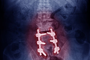
The benefit of walking for the management of the chronic low back pain patient is well-established in the literature and was previously discussed in the March issue. However, the patient with degenerative lumbar spinal stenosis (DLSS) has difficulty standing in corrected posture, let alone walking, making the prescription of walking challenging.
The common symptoms seen in DLSS are bilateral buttock and leg pain that increases with standing and walking, and abates rapidly with sitting and lumbar flexion. Patients over 70 years old are most affected and often demonstrate a wide-based gait for added stability.
The DLSS patient that presents with neurogenic claudication is a candidate for conservative care. The differential diagnosis includes vascular claudication from peripheral artery disease (affecting 26 percent of DLSS patients), radiculopathy, metabolic conditions (diabetes, hypothyroidism) and lumbopelvic hip pathomechanics.
 Clinical Tip: Have the patient sit tall on a stationary bike and cycle until buttock and/or leg symptoms occur. Instruct them to continue spinning and flex forward on the bike, placing the spine in flexion. Lessening of the pain while the patient continues to cycle indicates neurogenic claudication; no change or an increase in pain implies vascular claudication is present. A similar test can be done holding onto a shopping cart: first upright until pain starts, and then flexed to test for a decrease in symptoms.
Plain-film imaging is the most cost-effective assessment of lumbar degenerative changes. They are inexpensive, easy to obtain, give good assessment of the AP spinal canal diameter, and current high-speed systems have low levels of radiation. MRI is the diagnostic test of choice when additional information is needed concerning pathology, disc integrity, canal shape and adjacent soft tissues.
Clinical Tip: Radiographs of the lumbar spine need to be weight-bearing to assess the impact of axial load on the degenerative spine. For example, the extent of slippage in a degenerative spondylolisthesis may not appear in a recumbent film. Use an MRI center that performs upright or seated studies, and with both, consider flexion/extension studies.
Lumbopelvic hip imbalances and hip osteoarthritis can significantly impact DLSS. As the foundation of the spine, the hips and pelvis must be freely movable and stable to prevent compensatory movement patterns in the lumbar spine. Chiropractic adjustments, gluteal strengthening exercises and soft-tissue mobilizations are needed in to maintain optimum lumbopelvic hip mechanics.
Clinical Tip: Assess acetabular joint play and hip osteoarthritis. Restoring hip ROM and joint play, especially in internal rotation, is imperative for the DLSS patient.
Lumbar flexion is a position of relief for DLSS. Therefore, the active care progressions will favor a flexion-based approach as opposed to a neutral spine or extension-based approach. For example, strengthening the lumbar extensors is done from slight flexion, as in kneeling over a 45 cm physio ball and only extending to parallel, avoiding lumbar extension. Teaching your patients to auto correct their posture with a posterior pelvic tilt, creating lumbar spine flattening, is also necessary.
Clinical Tip: All intrinsic core stability exercises for the DLSS patient need to be performed in relative lumbar flexion by using a posterior pelvic tilt. Walking, too!
Stability and cardiovascular exercises need to be progressively loaded along with CMT and flexibility exercises. Recumbent or stationary bike is best for cardio activities and intrinsic core exercises that emphasize the transverse abdominus, internal oblique, pelvic floor and diaphragm. Remember, the rectus abdominus is not a core stabilizer.
Improving flexibility in the lumbar spine and legs is essential in this demographic. Carlo Ammendolia, DC, PhD, has developed a six-week exercise program associated with office visits twice a week that has shown significant improvement in functional indexes, including walking, in DLSS. The program uses post-isometric relaxation techniques to the hamstring, hip flexor and quadratus lumborum muscles to increase ROM. He also advocates flexion-distraction-style adjustments to improve lumbar flexion, soft-tissue compliance and flexibility.
These patients have a chronic condition and need to realize their stenosis needs to be managed. However, there is often an overlying tone of fear-avoidance behaviors and an emotional component leading to a negative health appraisal. The doctor needs to emphasize the functional goals of the treatment program and measure simple outcomes like walking distance and flexibility to shift the patient's focus from their limitations to their progress. As with any chronic pain patient, a referral for cognitive behavioral therapy may be needed.
Clinical Tip: Take the time to discuss functional goals to the patient and encourage them that exercise with discomfort does not mean additional disability. Dr. Ammendolia has also demonstrated that an individualized manual treatment program with self-management and behavior modification emphasizing functional tasks results in better outcomes and less negative emotional consequences.
In addition to central spinal stenosis, stenosis may occur unilaterally in the lateral recess of the spinal canal or even at the intervertebral foramen. This patient will have unilateral symptoms into the buttock or leg and responds well to soft-tissue mobilizations, flexibility and CMT – both flexion-distraction and diversified.
The DLSS patient is a challenge. They have chronic pain and often present with fear-avoidance behaviors and deconditioning. The research does not show significant long-term benefits with epidural injections, prescription medications or surgery. However, the research does show that the combination of addressing fear-avoidance behaviors, increasing flexibility and stability of the lumbopelvic hip region, and lifestyle guidance provides increased ability to walk and perform ADLs. For the doctor of chiropractic who is ready to roll up their sleeves and offer a multimodal approach, this is an opportune patient demographic.
Resources
- Ammendolia C, Chow N. Clinical outcomes for neurogenic claudication using a multimodal program for lumbar spinal stenosis: a retrospective study. J Manipulative Physiol Ther, 2015 Mar-Apr;38(3):188-94.
- Ammendolia C, et al. Comprehensive nonsurgical treatment versus self-directed care to improve walking ability in lumbar spinal stenosis: a randomized trial. Arch Phys Med Rehabil, 2018 Dec;99(12):2408-241.
- Lynch AD, Bove AM, Ammendolia C, et al. Individuals with lumbar spinal stenosis seek education and care focused on self-management - results of focus groups among participants enrolled in a randomized controlled trial. Spine, Aug 2018; 18(8):1303-1312.
- Schneider MJ, Ammendolia C, et al. Comparative clinical effectiveness of nonsurgical treatment methods in patients with lumbar spinal stenosis: a randomized clinical trial. JAMA Netw Open, 2019 Jan 4;2(1):e186828.
Click here for more information about Donald DeFabio, DC, DACBSP, DABCO.


