Patellofemoral pain syndrome is one of the most common gait-related disorders, affecting more than 25 percent of the running community.1 Despite the high prevalence, identifying the cause for this condition has been enigmatic.
Both of these conditions allow the patella to displace laterally into the lateral femoral condyle. As a result, various treatments were designed to correct faulty movement of the laterally displacing patella, with standard treatment programs emphasizing taping, soft-tissue mobilization and quadriceps strengthening. Unfortunately, these treatment protocols have been proven to be relatively ineffective in the management of chronic retropatellar pain.
Injury Mechanics
To get a better understanding of patellofemoral biomechanics in a weight-bearing environment, Powers, et al.,2 utilized dynamic MRI as subjects performed closed kinetic chain knee flexion. Surprisingly, these authors discovered the primary cause of lateral patellar displacement was not a shifting of the patella into the femoral condyle, but a shifting of the lateral aspect of the distal femur into the patella. Powers, et al., confirmed that contrary to popular belief, the patella does not shift into the stable lateral femoral condyle, but rather, the lateral femoral condyle rotates into the stable patella.
Recent CT and functional MRI evaluations support this observation, confirming the most likely cause of patellofemoral pain during weight-bearing is abnormal motion of the femur, not altered motion of the patella.3-4
Hip weakness has been cited as the most probable mechanism for the exaggerated internal femoral rotation. In a three-dimensional evaluation of runners with and without patellofemoral pain syndrome, Dierks, et al.,5 proved that runners with weak hip abductors have greater ranges of internal femoral rotation during stance phase, and the degree of rotation increases when runners become fatigued. Although the authors state that it is unclear whether hip weakness is a cause or an effect of patellofemoral pain syndrome, comprehensive conservative treatment should include exercises that target these specific muscles.
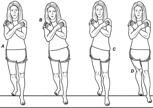
A clinically important test to help identify individuals in which hip weakness is affecting femoral rotation is the dynamic single-leg squat test. (See image) Developed by Crossley, et al.,6 this extremely useful test has excellent interrater reliability and a positive test has been correlated with delayed recruitment times in the hip abductor musculature. Simple pre- and post-treatment evaluations allow the practitioner to observe changes in motor recruitment patterns.

Orthotic Intervention
Besides hip weakness, another frequently cited cause of retropatellar pain is excessive pronation. Since too much pronation may allow the lower extremities to internally rotate through greater ranges of motion (displacing the lateral femoral condyle into the lateral patellar facet), controlling excessive pronation with orthotic intervention is a popular treatment protocol for managing patellofemoral pain syndrome.
In an extremely thorough randomized, controlled trial, Collins, et al.,7 confirmed that individuals with patellofemoral pain syndrome treated with prefabricated foot orthotics have significantly better outcomes compared to individuals treated with flat inserts. Although it is tempting to assume the orthotics are effective because they lessen pronation and hence internal rotation of the lateral femoral condyle into the patella, studies correlating improved outcomes with reduced ranges of pronation have found conflicting results.
Sutlive, et al.,8 report that people who pronate through small ranges of motion are more likely to have successful outcomes when treated with orthotics, while Vicenzino, et al.,9 note that individuals with greater midfoot mobility are more likely to have a favorable response to orthotic intervention. More recently, Crossley, et al.,10 found no correlation between excessive foot pronation and improved clinical outcomes following orthotic intervention. This is consistent with two- and three-dimensional studies confirming no correlation between patellofemoral pain and pronation.5,11
The main reason orthotic intervention produces such variable clinical outcomes when prescribed to alter pronation is most likely explained by arch-related variation in the location of the subtalar axis. As demonstrated by Williams, et al.,12 although people with low arches present with greater ranges of pronation during stance phase, they convert a much smaller percentage of frontal plane motion into tibial rotation. Conversely, individuals with high arches move through stance phase with smaller ranges of calcaneal eversion, but they convert a larger percentage of this motion into tibial rotation.
The end-result is that people with high and low arches move through the gait cycle with almost identical ranges of tibial rotation.12 This is consistent with a growing body of research showing a limited connection between arch height and patellofemoral pain syndrome.
This is not to say that orthotics should not be used in the management of this common disorder. Because orthotic intervention is associated with excellent outcomes in 25-40 percent of the patellofemoral pain population,7,13 the clinical challenge lies in identifying the individuals most likely to have favorable outcomes.
To address this issue, Collins, et al.,7 in their clinical prediction rules study, made the important observation that when individuals with patellofemoral pain report less discomfort when performing single-leg squats while wearing prefabricated orthotics, the potential that orthotic intervention will produce a marked reduction in symptoms after 12 weeks of regular use increases from 25 percent to 45 percent.
According to the authors, the patients most likely to respond to orthotic intervention typically claim the prefabricated devices make it easier to balance while performing the squat, and frequently report an increased ability to complete pain-free step-downs from a 20 cm high platform. Rather than functioning to reduce tibial rotation, orthotics may diminish retropatellar pain by enhancing proprioception, thereby improving motor control of the lower extremity.
Other Conservative Options for Management of PPS
The final consideration in the nonsurgical management of patellofemoral pain syndrome relates to the evaluation of alternate biomechanical factors that may increase the range of internal tibial rotation present during stance phase. The most common factor affecting tibial rotation is ankle equinus, in which an early heel lift causes the talus to adduct and plantarflex, thereby twisting the tibia inward.
It is also important to evaluate the integrity of the ankle ligaments following ankle sprain, because laxity of the anterior talofibular ligament significantly increases the transfer of calcaneal eversion into internal tibial rotation.14 Treatment of ankle laxity should include rock boards and closed kinetic chain exercises to stabilize the lower kinetic chain.
Manual therapies applied to the spine, hip, and knee should also be considered. In a recent randomized, controlled trial, van den Dolder and Roberts15 confirmed that transverse friction massage and patellar mobilization produce significant reductions in a functional step-down task compared to a control group. Spinal manipulation should also be considered, since it may improve neural drive to the quadriceps muscle.16
Finally, because running amplifies ground-reactive forces fivefold, overweight individuals should be encouraged to reduce body mass index, as even small reductions in weight may significantly lessen discomfort. In almost all situations, individuals with patellofemoral pain syndrome should walk and/or run with shorter stride lengths and consider switching to mid or forefoot strike patterns, which lessen the transfer of ground-reactive forces through the knee by as much as 50 percent.17
References
- Devereaux M, Lachman S. Patellofemoral arthralgia in athletes attending a sports injury clinic. Br J Sports Med, 1984;18:18-21.
- Powers C, Ward S, Fredericson M, et al. Patellofemoral kinematics during weight-bearing and non-weight-bearing knee extension in persons with lateral subluxation of the patella: a preliminary study. J Orthop Sports Phys Ther, 2003;33:677-685.
- Lin YF, Jan MH, Lin DH, Cheng CK. Different effects of femoral and tibial rotation on the dif-ferent measurements of patella tilting: An axial computed tomography study. J Orthop Surg Res, 2008;3:5.
- Souza R, Draper C, Fredericson M, et al. Femur rotation and patellofemoral joint kinematics: a weight-bearing magnetic resonance imaging analysis. J Orthop Sports Phys Ther, 2010;40:277-285.
- Dierks T, Manal K, Hamill J, Davis I. Proximal and distal influence on the hip and knee kinematics in runners with patellofemoral pain during a prolonged run. J Orthop Sports Phys Ther, 2008;38:448.
- Crossley K, Zhang W, Schache A, et al. Performance on the single-leg squat task indicates hip abductor muscle function. Am J Sports Med, 2011;39:866.
- Collins N, Crossley K, Beller E, et al. Foot orthoses and physiotherapy in the treatment of patellofemoral pain syndrome: a randomised clinical trial. Br Med J, 2008;337:a1735.
- Sutlive T, Mitchell S, Maxfield S, et al. Identification of individuals with patellofemoral pain whose symptoms improved after a combined program of foot orthosis use and modified activity: a preliminary investigation. Phys Ther, 2004;84:49-61.
- Vicenzino B, Collins N, Cleland J, McPoil T. A clinical prediction rule for identifying patients with patellofemoral pain who are likely to benefit from foot orthoses: a preliminary determination. Br J Sports Med, 2010;44:862-866.
- Crossley KM, Barton CJ, Menz HB. Clinical predictors of foot orthoses efficacy in patellofemoral pain syndrome. Med Sci Sports Exerc, 2010;42(suppl 5):Abstract 2611:S485.
- Hetsroni I, Finestone A, Milgrom C, et al. A prospective biomechanical study of the association between foot pronation and incidence of anterior knee pain among military recruits. J Bone Joint Surg, 2006;88-B:905-908.
- Williams D, McClay I, Hamill J, Buchanan T. Lower extremity kinematic and kinetic differences in runners with high and low arches. J Applied Biomech, 2001;17:153-163.
- Barton C, Menz H, Crossley K. Effects of prefabricated foot orthoses on pain and function in individuals with patellofemoral pain syndrome: a cohort study. Phys Ther in Sport, 2010 Oct 14.
- Sommer C, Hinterman B, Nigg B, vanderBogart A. Influence of ankle ligaments on tibial rotation: an in vitro study. Foot Ankle Int, 1996;17:79-84.
- van den Dolder PA, Roberts DL. Six sessions of manual therapy increase knee flexion and improve activity in people with anterior knee pain: a randomized controlled trial. Aust J Physiother, 2006;52:261-264.
- Suter E, McMoreland G, Herzog W, et al. Decrease in quadriceps inhibition after sacroiliac joint manipulation in patients with anterior knee pain. J Manip Phys Ther, 1999;22:149-153.
- Cavanagh P, Lafortune M. Ground reaction forces in dis-tance running. J Biomech,1980;13:397-406.
Click here for more information about Thomas Michaud, DC.


