


Patients may present with a combination of structural dysfunction due to ligament-capsular-discal derangement and/or myofascial involvement due to trigger points of the masseter, temporalis and pterygoids. TMJ syndrome may occur as a result of the cervical acceleration/deceleration syndrome, as well as other structural problems such as posture, stress with or without bruxism, malocclusion, leg-length discrepancy, and blunt trauma.
A full discussion of the TMJ syndrome is beyond the scope of this paper, but when a patient presents with the above signs and symptoms, the treating doctor should have a high index of suspicion for TMJ syndrome. Diagnostic imaging of the TMJ includes radiographs of the TMJ, open and closed mouth, as well as panorex views. MRI may be helpful to diagnose structural abnormalities such as torn temporomandibular ligaments or anteriorly displaced disc.
A cost effective, noninvasive, risk free, imaging tool that may be utilized is thermography. While x-ray and MRI can show structural/morphological aspects of the TMJ syndrome, thermography can show physiological information with reference to dysautonomia associated with TMJ as well as myofascial pain dysfunction (MPD) and trigger points. Thermography is recommended as one of the diagnostic tools useful for TMJ syndrome, by the American Academy of Head, Neck, Fascial Pain and TMJ Orthopedics published in a position paper in Cranio: The Journal of Craniomandibular Practice, in January 1990.
In a paper published in the Clinical Journal of Pain in 1987, by Weinstein et al., a TMJ protocol was described for thermographic imaging of the facial area in patients with TMJ syndrome. In their study of 30 patients with TMJ symptoms, they found a 95 percent correlation between anatomical distribution of symptoms and thermographic findings. Their TMJ protocol included using a half degree level of sensitivity and a one-half degree delta T (temperature differential) from right to left. They recommended a triplicate series of facial views including; frontal, right and left lateral facials, trigger point views of the anterior cervical spine, posterior cervical spine, and interscapular region. Weinstein's criteria finds a positive study when:
- affected joint is 1.0 degree centigrade warmer than the opposite side in comparison to the ambient room temperature;
- each affected masseter is 1.0 degree centigrade below ambient room temperature;
- location of the pain pattern matches the pain diagram.
In a study published by Pamela Steed, D.D.S., and published in the Journal of Craniomandibular Practice, she found thermography to be very useful in the diagnosis and management of TMJ syndrome. She found a 94.5 percent sensitivity using thermography for TMJ syndrome, which correlates well with the Weinstein Study. After treatment, thermography was able to assess the successful outcome of treatment in 80 percent of the cases. She was also able to find a pattern that could differentiate acute vs. chronic TMJ dysfunction.
In another study published by Weinstein, they documented normal temperature differences in the face (see Table 1 and 2). They also found that 0.5 degrees Celsius difference from right to left of the face had a 96 percent confidence level for underlying pathology.
Table I | |||
Mean Skin Surface Temperature Value Differences: Male versus Female | |||
| Muscle | Mean Difference Male vs. Female (oC) | Mean Male (oC) | Mean Female (oC) |
| L. temporalis | 0.7 | 37.18 | 37.12 |
| R. temporalis | 0.5 | 37.24 | 37.18 |
| L. frontalis | 0.1 | 37.07 | 37.18 |
| R. frontalis | - | 37.11 | 37.15 |
| L. peripunctal area | 0.2 | 38.4 | 38.19 |
| R. peripunctal area | 0.22 | 38.38 | 38.15 |
| L. front masseter | 0.27 | 35.96 | 35.69 |
| R. front masseter | 0.36 | 36 | 35.64 |
| L. mentum | 0.06 | 36.93 | 36.99 |
| R. mentum | 0.07 | 36.94 | 37 |
| L. sup. temporal artery | 0.02 | 37.99 | 37.77 |
| R. sup. temporal artery | 0.21 | 38 | 37.79 |
| L. temporomandibular joint | 0.4 | 37.12 | 36.72 |
| R. temporomandibular joint | 0.6 | 37.09 | 36.7 |
| L. lateral masseter | 0.39 | 36.4 | 36.02 |
| R. lateral masseter | 0.41 | 36.44 | 36.02 |
Table II | |
Mean oC | |
| 1. Right Temporalis Muscle | 37.21 |
| 2. Right Frontalis Muscle | 37.13 |
| 3. Left Frontalis Muscle | 37.12 |
| 4. Left Temporalis Muscle | 37.15 |
| 5. Right Peripunctal Area | 38.28 |
| 6. Left Peripunctal Area | 38.31 |
| 7. Right Masseter Muscle | 35.84 |
| (Frontal) | |
| 8. Right Masseter Muscle | 35.84 |
| (Frontal) | |
| 9. Right Mentum | 36.97 |
| 10. Left Mentum | 36.98 |
| 11. Right Superficial Temporal Artery | 37.91 |
| 12. Right Temporomandibular Joint | 36.92 |
| 13. Right Masseter Muscle (Lateral) | 36.25 |
| 14. Left Superficial Temporal Artery | 37.89 |
| 15. Left Temporomandibular Joint | 36.93 |
| 16. Left Masseter Muscle (Lateral) | 36.23 |


Dr. S.A. & G. Weinstein
Summary of TMJ findings with infrared imaging:
- Focal hyperthermia over the TMJ.
- Focal hot spots consistent with trigger points at the paracervical, interscapular, suprascapular region, and the masseter region.
- Decreased thermal emission over the masseter muscle area.
- Increased thermal emission over the temporalis muscle area.
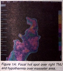
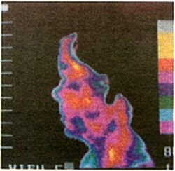
Figure 1B: No hot spot over TMJ and no cooling over masseter compared to symptomatic right side.
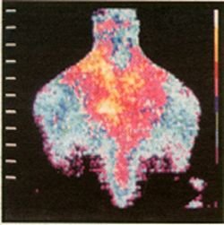
Figure 1C: Trigger points in the neck, levator scapulai, and rhomboids often seen with TJM sydrome.
Conclusion
Thermography is a useful diagnostic imaging tool available to the treating doctor to differentially diagnose this multifaceted complex syndrome from other conditions with similar symptoms. The TMJ syndrome has a distinct thermal pattern. Thermography is also useful to help follow the patient's response to treatment.
Reference:
- Weinstein SA: A protocol for the identification of temporomandibular joint disorders by standardized computerized electronic thermography. Clinical Journal of Pain, 3:107-112.
- Weinstein SA: Thermophysiologic anthropometry of the face in Homosapiens. Journal of Craniomandibular Practice, 8(3):252-255, 1990.
- Weinstein SA: Facial thermography, basis, protocol, clinical value. Journal of Craniomandibular Practice, 9(3):201-211, July 1991.
- Chapman GE: TMJ Dysfunction and Thermal Imaging. Chiropractic Products, pp 98-100, Dec. 1988.
- Finney JW, Holt CR, Pearce K: Thermographic diagnosis of TMJ. Postgraduate Medicine, pp 93-95, March 1986.
- Steed P: Utilization of contact liquid crystal thermography in the evaluation of temporomandibular dysfunction. Journal of Craniomandibular Practice, 9(2):121-127, April 1991.
- BenEliyahu DJ: Thermography in clinical chiropractic practice. ACA Journal of Chiropractic, pp 64-65, August 1989.
David BenEliyahu, D.C., CCSP, DNBCT
Selden, New York