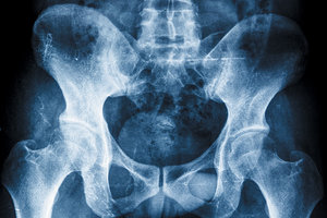
Treatment of pelvic dysfunction that is effective and lasts beyond our patients walking out to the parking lot to their car requires observing and thinking about tri-planar movement of the pelvis.
The pelvis, as well as our entire body, needs to have as close to symmetrical muscle flexibility, strength and length as possible. Asymmetries in muscle strength, length and endurance with agonists and antagonists can eventually create dysfunction in movement and alignment patterns. Our rehabilitative goals need to include addressing these asymmetries effectively.1
We are all born with asymmetries that the body deals with effectively for the most part. However, the body needs our help when those asymmetries form dysfunctional myokinematic patterns that lead to pain, decreased performance and ultimately, premature degenerative changes.
Common Asymmetries
Let's explore some of the many asymmetries we as humans possess and need to integrate and balance every minute of every day. For starters, we have a left- and right-sided portion of our brain, with the left side associated with motor skills and analytical thought. The right side is associated more with abstract thought and creativity. The autonomic nervous system, with sympathetic and parasympathetic portions, can often be out of balance and asymmetrical. Would you agree that in this day and age, there is too much stimulation or "tone" on the sympathetic or fight-or-flight side?
 The lymph system is also asymmetrical, with drainage on the right side clearing the right arm and chest, and the rest of the body being drained on the left. What's more, most of us are right-hand dominate over the left. And if you take a picture of your face, split it in half, use a mirror image and combine right to right and left to left sides, you may not recognize yourself!
One of the most significant asymmetries our bodies has to integrate involves the diaphragm or "Big D" and respiration, which is key when considering effective treatment of musculoskeletal function.
There are two leaflets of the diaphragm, with the right side having bigger and more muscular "crura." The left side is smaller, has less muscular crura and attaches 1 to 1 ½ lumbar vertebra higher than the right. On the right side of the abdominal cavity is the liver, which helps "dome" the diaphragm on that side. There are also three lobes of lung on the right side, as opposed to two lobes on the left, with the heart and aorta orienting more toward the left. A small muscle in the anterior chest wall called the transversus thoracis or triangularis sterni helps balance this asymmetry.2
Importance of the Diaphragm
The diaphragm may be the most important muscle in the body. Obviously, it is the key muscle for breathing, but it also must stabilize the lumbar spine and torso. Breathing, stabilizing and even walking are just some of the functions connected to Big "D."
Typically, the left side of the chest wall "flares" because of inactivation of the anterior lateral abdominal muscles, combined with a flat or over-activated diaphragm on the left side. Remember, it doesn't dome like the right side does, and full exhalation does not commonly occur on our left side unless we are trained to do so.
The inability to exhale completely and coordinate the diaphragm with the abdominal wall results in a faulty "Zone of Apposition" (ZOA), causing pelvic misalignment as well as thoracic misalignment. (Refer to part 3 of my recent series on how to "Breathe Well and Breathe Often" in the Sept. 9, 2012 issue of DC.) This connection and potential asymmetry of the diaphragm have a profound effect on lumbar spine alignment!
Understanding ZOA is fundamental to understanding the close relationship with the diaphragm, as well as the sequence of muscles that create a "polyarticular chain" that affects all neuromuscular skeletal movement and function. To understand pelvic dysfunction and how to restore balanced biomechanics, we need to look at groups of interconnected muscles and how they interact in three planes of movement.
Ron Hruska, MPA, PT, director of the Postural Restoration Institute, describes the "anterior interior chain" (AIC) composed of muscles that form a polyarticular connection, starting with attachments to the costal cartilage and bone of ribs 7 through 12; then terminating at the lateral patella, head of the fibula and lateral condyle of the tibia.
These two tracts of muscles, left and right side, are comprised of the diaphragm to psoas muscle and then with the iliacus, TFL, biceps femoris and vastus lateralis, combining a chain of muscles that have significant influence on balanced pelvic motion, breathing and gait.3
(This left and right AIC has a profound effect on another polyarticular set of muscles called the brachial chain and will be described in a future article.)
Tone and Asymmetry
"Tone," either too much or too little, can have a profound influence on an asymmetry and whether that asymmetry will affect us in terms of function, athletic performance or chronic pain in the spine, pelvis or an extremity. It is important to remember that asymmetries, anatomical or neurological, are not usually a problem or issue because our bodies have a system of homeostasis that helps us adapt, balance and adjust to those asymmetries.
It is only when those asymmetries become too excessive and we are unable to restore balance that they are a problem. One of the goals, as James Anderson, MPT, PRC, states in his course on myokinematics, is that these patients with too much tone just want to relax!
The side that usually has too much tone for most human beings is the left anterior interior chain, unless you are born with a liver and three lobes of lung on the left side. ("Situs inversus" is a rare condition in which the organs are actually flip-flopped from their natural position.) The reasons for this are many, but let's just start with the diaphragm being flatter or less domed than the right side.
Remember that when the diaphragm contracts, the central tendon drops and the diaphragm flattens to create negative pressure in the chest cavity, so the lungs fill with air. Typically, most people have a dominate left anterior chain pattern because if the diaphragm doesn't completely relax, neither will the rest of those muscles mentioned in the left polyarticular chain. (In fact, the muscle fibers of the diaphragm and the psoas are so closely interrelated that upon dissection, it is nearly impossible to distinguish between the two.)3
This excess tone or inability to relax the left anterior chain of muscles has many consequences anatomically. Typically, there is an "orientation" of the sacral region to the right with an anterior tilt and flexion of the hip on the left side, if you are viewing from above in the transverse plane. In addition, with the above-mentioned rib flare, especially on the left, the thorax tends to rotate to the left, creating the opportunity for scoliosis, rotational / compressive forces to the discs, and excessive stress to the facet joints of the thoracolumbar spine because of a rotational / extension alignment and movement pattern.1
In Part 2 of this article, I will describe specific muscles and the myokinematic pelvic patterns they influence. I will also describe the importance of the diaphragm and breathing with every one of our patients. And ultimately in this series on the pelvis, I will describe corrective treatment strategies that help evolve our patients from purely passive and dependent care to being more active and independent participants in their well-being!
References
- Myokinematic restoration. Postural Restoration Institute home study course, pages 1, 8.
- Anderson J. Postural respiration seminar, May 18-19, 2012. Lecture notes, page 11.
- Postural respiration seminar, May 18-19, 2012. Lecture notes: Brachial Chain and Anterior Interior Chain (page VI); the Left Anterior Chain Pattern (page vii); Zone of Apposition by Ron Hruska, MPA, PT (page viii).
Dr. Robert "Skip" George practices in La Jolla, Calif., where he integrates chiropractic, rehabilitation and sports performance training. He is a certified Functional Movement Screen instructor and has lectured nationally on subjects related to the chiropractic profession. He can be contacted with questions and comments at
.


