Hypomobility of the first rib has been suggested as a possible mechanism causing thoracic outlet syndromes (TOS).
(1) The stellate ganglion lies adjacent to the first costotransverse joint.

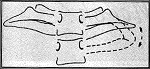
A schematic representation of the normal and restricted first rib movement.
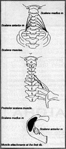
2. The first costotransverse joint is the only level that lacks ligamentous support superiorly and is dependent on motion of the scalene muscles for support and respiratory motion, making this joint susceptible to biomechanical disorders that affect the lower cervical spine and musculature. The anterior middle and posterior scalene muscles originate at the transverse processes of the C3-C7 vertebral segments and insert on the first and second ribs.

Origin and insertion of the scalenes
Patients with lower cervical subluxations and fixations may also have associated symptoms which involve the thoracic outlet due to this intimate relationship between the first rib and the scalene muscles.
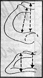
The motion of the first rib can be evaluated by palpating the first rib under the clavicle during respiration or radiographically by performing inspiration and expiration views of the first rib. Respiratory movement of the first rib can be measured from an AP view of the cervical thoracic junction by measuring the distance between the anterior and posterior margin of the first rib during maximal inhalation and exhalation.

Landmarks used in the measurement of first rib excursions during respiration.
The distance between the anterior and posterior portion of the first rib diminishes with inhalation, since the anterior aspect of the rib elevates during inhalation and descends with exhalation.
Along with adjusting the cervical spine, mobilization of the first rib should be performed if the first rib demonstrates fixation. The technique generally suggested is isometric exercises. The patient's head should be pushed against the palm of the painful side, moving forward, to the side, and backward, thus isometrically contracting the anterior, middle, and posterior scalenes. Each movement should last at least one second, with a short pause between positions. This series of pushing movements should be repeated ten times. Motion palpation of the first rib can then be performed to gauge the patient's progress.
Once the first rib is able to move freely with respiration, often the brachialgia can be alleviated.
References
Lindgre K, Leino E: Subluxation of the first rib, a possible thoracic outlet syndrome mechanism. Arch Phys Med Rehabil., 69:692-695, 1988.
Deborah Pate, D.C.
San Diego, California
Click here for more information about Deborah Pate, DC, DACBR.


