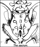
Sacral restrictions have to be differentiated from restrictions of the ilio-sacral joint. In osteopathy, most techniques separate the sacroiliac and ilio-sacral joints.
In my opinion, the best way to look at the sacrum is to assess it embryologically, as five segments. We are born with our sacral segments unfused. Paul Chauffour [pioneer of the "mechanical link" technique] first introduced me to this concept. One way to visualize or model this is to see the sacrum as having intraosseous restrictions (see www.chiroweb.com/archives/19/23/12.html). This model implies that bones themselves should have a certain amount of flexibility, and that a restriction within the matrix of the bone itself is a clinically important form of subluxation. Bones are not dead tissue made only of calcium, but live structures with an active blood supply and multiple physiological functions. The assumption here is that the sacrum, like all live bones, has give, and should be somewhat supple. Don't visualize pickled cadaver bone; visualize live bone, full of innate intelligence. If this conceptual leap is a bit much for you, imagine the most restricted and tender spot on the sacrum as the best leverage point for releasing the sacrum as a whole.
As usual, the first step is to identify the lesion. Most of my sacral work is done with the patient prone. Use "listening," by placing your hand over the sacrum, with your palm accommodating the kyphotic surface of the bone. Where are you attracted? You can begin by dividing into quadrants: upper right; lower right; lower left; and upper left. To which quadrant are you attracted? Within the quadrant, are there a specific spot(s), is there a specific spot (or spots) that attracts you? The lesion could be midline, or it could be off to one side.
Next, palpate. You can use your thumb pad, finger pads or thenar pad. Begin gently with "initial response testing" (see www.chiroweb.com/archives/19/16/12.html). You are looking to differentiate "give" versus "nongive": a sense of mobility versus rigidity. You are touching a small spot within a large bone, and not really looking for gross range of motion, just that sense of stiffness versus mobility. Once you have found an area of increased stiffness, press a little harder - enough to elicit tenderness from the patient. Significant lesions have tenderness and restriction.
You've found a place; let's say it's the second sacral segment on the left: LS2. Get specific and map the 3-D restriction. Think of this spot as analogous to a vertebral segment. Does it resist flexion or extension? Does it resist torquing or rotation around an anterior-posterior axis to the left or right? Does it resist a sidebending or lateral motion to the left or right? You can test these one at a time, or just listen, and get the whole 3-D assessment in one moment.
Another possibility is that the segment is stuck, compressed or expanded along a vertical or horizontal line. George Roth [developer of the "matrix repatterning" technique], with his focus of trauma stored in the bones, introduced this idea to me. Imagine the forces that go into the sacrum when someone falls on his or her hip. Can you visualize a slightly oblique horizontal compression line, along which a part of the sacrum is compressed? Test this by decompressing along this line, using both thumbs. How about a fall on the buttock, with a more vertical line of compression? Imagine the torque applied to the sacrum in a twisting motion, accompanied by sudden muscle spasms, as in when you lose your balance and catch yourself. If the force of the trauma were to stretch the bony sacral fibers, you might find an expanded lesion that needs testing and correcting via compression from both sides, or from top to bottom. These lines of compression or expansion may be straight and horizontal or vertical; or oblique, with a spiraling component. Listening is a great tool to assess this with, but step-by-step testing in various directions also will provide the same information. You have a specific spot to test from, based on your listening and palpation. Test here vertically and horizontally, in compression and expansion. Once you feel a restricted direction, fine-tune for the oblique angles. In your correction, you'll take care of any spirals.
Sacral Correction
How do I correct the lesion? The patient is prone. My usual correction here uses recoil ("engage-release") or "engage, listen, follow" (ELF) techniques (see www.chiroweb.com/archives/19/18/07.html). On a hard, bony structure such as the sacrum, I'll usually start with ELF, and include a couple of recoils during the process. I can do an ELF correction in a direct (toward the barrier), or indirect (toward ease) direction. I usually start directly, going toward the 3-D barrier. I switch to indirect if I don't get a complete release, or if I feel I am up against a brick wall, with absolutely no "give."
If I feel the involved area includes the ilium or the parasacral soft tissues, I'll often use a long-lever technique, side-lying. I could side-lie the patient and use an HVLA thrust, focusing on the specific sacral segment. More often, I'll side-lie the patient, set him or her up as if I were going to thrust, stretching across the SI joint; then I instruct the patient to push the bent leg upward isometrically toward the ceiling against the resistance of my thigh or hand. Lastly, the patient must relax as my contact hand takes the sacral segment into further motion. This is a muscle-energy variation on side-posture adjusting. As in all muscle-energy techniques, we use exact positioning of the long lever to fine-tune the sense of the barrier. We modify the degree of hip flexion, extension and adduction of the thigh to find our ideal position. It allows me to stretch and release the tissues all the way from the hip through the ilium and into the sacrum.
The Sacro-Tuberous Ligament
Another significant structure that affects the sacrum is the sacrotuberous ligament. Most of us address the piriformis, but (except for Logan Basic practitioners) few of us assess and correct the sacrotuberous ligament directly. A lesion here is found the usual way, expressed as tenderness and restriction. I'll palpate from inferior upward and from medial to lateral, deep in the buttock, directly contacting this ligamentous band. Ligaments can palpate as short and tight, and can lengthen or release immediately. It is the same phenomena we find with any adjustment of any joint, in which the joint capsule ligaments go from a stiff state to a more mobile state. I'll correct the sacrotuberous ligament directly by using ELF to release it in the direction of restriction.
My next article will address the coccyx, completing our pelvic section. I trust you will find that discussion useful for your practice, as well. No one piece stands on its own; it integrates as a whole. Fix what you find, and let innate do its healing work.
Resources
- Paul Chauffour. Mechanical Link, North Atlantic Press, 2002.
- George Roth. Matrix repatterning courses.
- Marc Heller. Framework courses and articles.
Click here for more information about Marc Heller, DC.


