


|
|
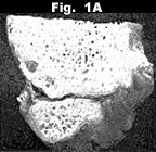
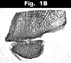
| Figures 1A and 1B: Radiograph and photograph of an anatomic specimen. Note the fine cystic changes in the region of the surface of the tibiale externum. | |
|
|
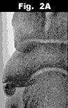
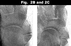
| Figures 2A, 2B and 2C: These are examples of a "painful os tibiale externum." A is a 16-year-old with fine cysts in the coalescing surfaces; B is a 35-year-old with degenerative changes at the adjacent margins; and C is a 50-year-old with relatively mild changes of the adjacent margins. | |
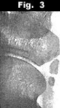 Figure 3: Bipartite os tibiale externum in a 12-year-old. Note the appearance of the ossicles in the tendon.
 |
|
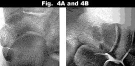
| Figures 4A and 4B: AP (A) and lateral (B) view of a bipartite os tibiale externum. Note the smaller ossicle is barely visible on the AP view and is under the head of the talus in this view. |
The ossicle may consist essentially of connective or chondroid tissue, and will undergo enchondral ossification with the maturation of the skeleton. The ossicle is situated in the tendon of the posterior tibial muscle that also inserts on the navicular tuberosity. In the first decade of life, the tendon of the posterior tibial muscle forms the connection between this accessory ossicle and the navicular bone. Later, the ossicle grows more out of the tendon. The interspace between the two bones is filled in with connective tissue or with chondroid and fibrocartilage. No joint is formed. So what's the point? The os tibiale externum can often be associated with symptoms because of its exposed position, and often after sprains to the ankle and foot.
In cases of painful os tibiale externum, one may see small cystic lesions, calcifications and deformities due to arthrosis or dislocation. Fractures of the navicular bone are relatively uncommon. The most frequent is an avulsion fracture of the dorsal surface of the talonavicular joint, which is difficult to identify except on a true lateral view. Next in frequency are fractures involving the tuberosity on the medial aspect of the navicular bone. The tuberosity serves as the major insertion for the posterior tibial tendon. Fractures of the navicular often are associated with fracture of the cuboid.
If the injury is a simple sprain and the patient has an os tibiale externum, take an x-ray of the opposite foot; hopefully, the asymptomatic foot and the answer between fracture and ossicle should not be difficult to discern.
Deborah Pate,DC,DACBR
San Diego, California
![]()
Click here for more information about Deborah Pate, DC, DACBR.