


The lucent cleft sign, mainly seen with the spine in extension, can be visualized in the cervical and lumbar regions. It represents gas, most of which is probably nitrogen, in the region where the disc has partially torn away from the vertebral body endplate.
The traumatic avulsion of the disc from the endplate with negative routine radiographs was first reported in 1948 by Taylor and Blackwood. They described a fatal hyperextension neck sprain in a patient who presented just prior to death with severe cervical cord damage and negative x-rays. On extension of the cervical spine at the autopsy, complete detachment of the intervertebral disc from the vertebral body endplate was discovered, along with a rupture of the anterior longitudinal ligament. Apparently there was an acceleration which had caused the injury, but it stopped abruptly. Spontaneous reduction occurred to such a degree that subsequent radiographs appeared normal.
Vacuum clefts are well-known in syndesmoses joints under stress. The stress suddenly widens the joints space, reducing the pressure to below vapor pressure of the gases, dissolved in the synovial fluid cartilaginous joint surfaces. These gases diffuse into and fill the expanded joint space, which appears lucent on radiography.
With the injured cervical spine in extension, the potential space where the disc is partially avulsed from the cartilaginous endplate is pulled apart, much like the joint under stress described above and results similarly in a lucent cleft (see Figure 1). The lucent clefts in the case of a traumatic injury will be noted within hours of the acute trauma and may persist unchanged from two months to five years. There are no potential spaces within a normal disc, nor at the junction with the endplate, and therefore no lucent cleft are seen in the extension lateral view of the normal cervical spine.
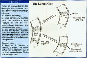
In the case of complete avulsion of the disc and rupture of the anterior longitudinal ligament, a lucent cleft is unlikely to be visualized due to the associated local hemorrhage which fills the cleft with blood. With less severe injury, the lucent cleft is visualized on the extension view and is a definite indication of avulsion.
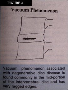
The other vacuum sign demonstrated in the intervertebral disc is described as the vacuum phenomenon, and is associated with disc degeneration. The vacuum sign of disc degeneration has ragged borders, often is seen involving the middle of the intervertebral disc and is indicative of disc desiccation. This is not a sign of acute trauma and is commonly seen in patients with moderate-to-severe degenerative disc disease (see Figure 2).
The vacuum cleft sign seen in patients with a hyperextension/hyperflexion injury is a very significant radiographic finding which confirms the existence of an avulsion injury to the intervertebral disc. This injury should be managed with care, as continued extension may aggravate the injury.
Deborah Pate, DC, DACBR
San Diego, California
Click here for more information about Deborah Pate, DC, DACBR.