


Patients may occasionally ask you to take a chest x-ray. Often the chest x-ray is a pre-employment requirement. If you choose to take chest x-rays for your patient, please be aware of the technical requirements.
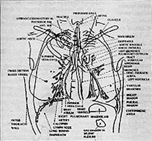
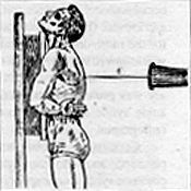
PA Chest:
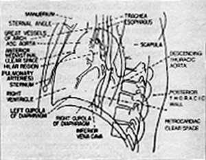
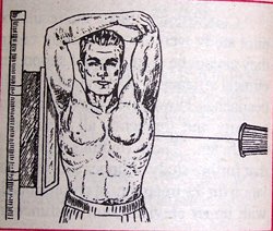
Lateral Chest:
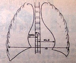
The Cardiothoracic Ratio:
This measurement is performed to determine the size of the heart and a screening for heart enlargement.
MRD = maximum transverse diameter of the right side of the heart, which is a line drawn from the midline of the spine to the most distant point of the right cardiac margin.
ML= midline of the spine
MLD = maximum transverse diameter of the left side of the heart, which is a line drawn from the midline of the spine to the most distant point of the left cardiac margin
ID= greatest internal diameter of the thorax
TD = MRD + MLD
cardiothoracic ration = TD/ID
Deborah Pate, DC, DACBR
San Diego, California
Click here for more information about Deborah Pate, DC, DACBR.