


The subluxation is an accepted term used to describe joint malalignment without complete dislocation. Unfortunately, this finding is often overlooked by medical radiologists. We all know that subluxation includes more than just a joint displacement.
Subluxations associated with trauma consists of minor forward or backward displacement of one vertebra on another. Harris lists subluxation under stable cervical injuries of the spine.1 According to Jackson2 subluxations can be noted on lateral views of the cervical spine and many times are accentuated in flexion and extension views. The backward subluxations (retrolisthesis subluxations) are present when the posterior margin of the vertebral body which is displaced posteriorly is seen to lie behind the posterior margin of the adjacent distal vertebral body. The superior facets of the lower vertebra can be displaced forward enough to encroach on the intervertebral foramina (Figure 1).
Forward displacement of one vertebral body on the vertebral body below, indicates rupture of the posterior ligamentous structures and joint capsule.2 Most orthopedic surgeons recognized subluxations as instability of the posterior ligament complex. In Cheshire's study on the stability of the cervical spine, he reported a 21 percent incidence of delayed instability was associated with anterior subluxations, as compared to four percent of all types of cervical injuries.3 Delayed instability is defined as the presence of "abnormal mobility between any pair of vertebrae, with or without pain or other clinical manifestation, when lateral x-rays of the cervical spine are taken in flexion and extension at the conclusion of conservative treatment."3
Subluxation as defined by Harris is the result of a purely soft- tissue injury consisting of disruption of the posterior ligament complex (the supraspinous, interspinous, and posterior longitudinal ligament, the capsule of the interfacetal joints and ligamentum flavum), and in addition, possible tear of the posterior portion of the intervertebral disc (Figure 2).
Radiographically, a subluxation level can be found by evaluating the interspinous spaces in the neural lateral projection. Fielding and Hawkins have referred to a widening of the interspinous space as fanning, indicating a subluxation.4 Other radiographic signs are upward displacement of the superior facets and forward with respect to the contiguous inferior facets. The disc space is widened posteriorly and narrowed anteriorly as a result of forward displacement of the involved vertebral body on its anterior inferior corner. For this reason, kinetic studies of the cervical spine are highly recommended when a subluxation is suspected.
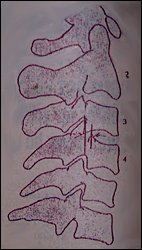
Figure 1: Backward subluxation or retrolisthesis subluxation
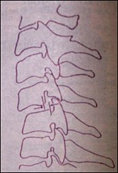
Figure 2: Forward or anterolisthesis subluxation.
References
- Felson B: Roentgenology of Fractures and Dislocations. Harris J: Chapter on Spine.
- Jackson R: The Cervical Syndromes, pp 180-187, Charles Thomas, 1971.
- Cheshire, DJ: The stability of the cervical spine following the conservative treatment of fractures and fracture-dislocations. Paraplegia, 7:193-203, 1969.
- Fielding W, Hawkins R: Roentgenographic diagnosis of the injured neck. Chapter 7, AAOS Instructional Course Lectures, 149-169, 1988.
Deborah Pate, D.C., DACBR
San Diego, California
Click here for more information about Deborah Pate, DC, DACBR.