


Injuries to the pediatric elbow can be very difficult to detect radiographically when one is not aware of the different ossification centers that can be present. A center of ossification appears in the head of the radius in the fifth and sixth year of life and fuses with the radial shaft at the age of sixteen to nineteen.
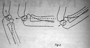
One of the most common problems in young children is the anterior subluxation of the radial head. Many times just the history can be sufficient enough to give the examiner a preliminary diagnosis. When a child running beside an adult, stumbles; the adult jerks the child upright by a tug on his hand to prevent the child from falling. Usually this injury will cause pain and the child will cry for a few minutes and then will continue about his play, but will not move the injured arm. This injury has been named the nursemaid's elbow. In young children the annular ligament is easily stretched, which will allow the radial head to slip from its fascial containment. (Fig 1) Upon examination, the child will not allow the examiner to supinate the hand. X-rays are generally negative for osseous pathology. The relationship between the capitellum and distal radius may be slightly malaligned. (Fig 2)
Treatment for this injury is simply closed reduction. Reduction can be performed by gripping the elbow gently with one hand with the thumb over the radiohumeral joint and with the opposite hand rotating the forearm, first into full pronation, then into full supination. Reduction will be perceived as a click by the stabilizing hand at the end of supination. In ten to fifteen minutes the child will be using his arm again, provided the reduction has been accomplished.
Fig 1 (A and B) Humeral condylar epiphysis.
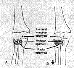
Fig 2.
Click here for more information about Deborah Pate, DC, DACBR.