Chiropractic treatment for extremity dysfunctions has a long history.1 Extremity dysfunction has also been correlated with muscle weakness extensively in the research literature.
Extremity function is an interaction of many local and remote structures, depending on proper nerve receptor stimulation, blood vascular supply and other factors. The many structures that contribute to extremity movement compound the diagnostic problem, and there may be more than one problem.18
In chiropractic, Goodheart first introduced methods for detecting extremity dysfunctions with the manual muscle test (MMT) for the shoulder in 1964, the foot in 1973, the wrist in 1974, the elbow in 1976 and the ankle in 1977.19 Today, the chiropractic approach to extremity conditions (encouraged by the AK demonstration of the complex interactions in this system) is multi-modal; it considers the body from the feet to the cranium. Many think of the sacrum as the foundation of the spine, but as Gillet and Liekens point out, the ischia are its base when sitting and the feet when standing.20 The importance of the intrinsic and extrinsic muscles of the feet, and their primary role in foot dysfunctions, has been described by many authors.
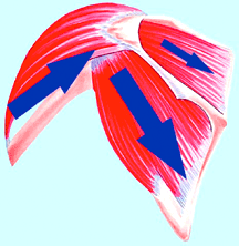
 The glenohumeral joint is best considered for evaluation and treatment as a muscular socket, not a bony socket. Its muscles protect the joint, ligaments, and bursa. Each of these muscles can be specifically assessed with the MMT in order to guide treatment to the dysfunctional component in the shoulder complex.
The glenohumeral joint, for instance, is best considered for evaluation and treatment as a muscular socket, not a bony socket. The only skeletal attachment of the shoulder-arm complex to the body is the sternoclavicular joint, which provides inadequate stability. Stability and control are basically up to the muscles.
As noted throughout the MMT literature, a muscle must function from a stable base to test strong. For example, stability of the clavicle and/or scapula is essential in shoulder muscle function, and if during your examination of the shoulder there is weakness on the shoulder MMT, re-evaluate the test by stabilizing the clavicle or scapula. If the pectoralis (clavicular division) tests weak, stabilize the clavicle and re-test the muscle. If it then tests strong, test for subluxations of the sternoclavicular and acromioclavicular articulations. When lack of clavicular or scapular stability is causing a shoulder muscle to test weak, determining the reason for the instability goes a long way toward correcting shoulder dysfunction.
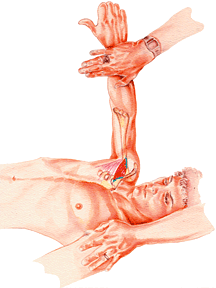
This is an illustration of the AK challenge and therapy localization methods. These tools, along with functional evaluation of muscles, provide valuable additional tools for the chiropractor in the evaluation of the total extremity joint complex.
 The pectoralis (clavicular division) manual muscle test.
Ideally, correction is directed toward primary factors. Primary factors can be found by applying numerous examination techniques at once or in succession to determine what eliminates dysfunction. In the example above, if the pectoralis (clavicular division) muscle tests weak and challenge to the clavicle or some other bone causes it to test strong, the subluxation is primary. Although the pectoralis (clavicular division) could probably be strengthened by stimulating the neurolymphatic or neurovascular or acupuncture reflexes, or with other types of rehabilitative exercises, it is likely it would immediately lose its correction with gait or some other structural stress to the clavicle. If so, the primary cause of the pectoralis (clavicular division) weakness appears to be the result of improper stimulation to receptors by the subluxated joint. It might be due to the muscle vainly trying to stabilize the clavicle or some other bone in subluxation, and weakening because of its inability to do so.
It was suggested as far back as the turn of the century that ligamento-muscular reflexes exist from sensory receptors in ligaments to muscles that modify the load imposed on the ligament and joint. Goodheart first discussed the law of the ligaments in 1973.19 He found that pressure applied to the ends of ligaments toward the belly of the ligament tightens it. The opposite force will elongate the ligament.
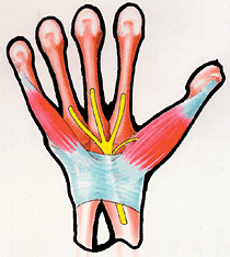
 Peripheral nerve entrapment of the median nerve at the carpal tunnel or elsewhere in the arm or neck can be differentially diagnosed using the MMT.
It has been shown that ligamento-muscular reflexes exist in most extremity joints.21-23 The ligaments associated with each extremity are richly endowed with afferents that produce reflex activation of the many muscles associated with the extremity's movement. The muscles, therefore, are a major component in maintaining the stability of the extremity's ligaments, bursae and capsules.24-25
Goodheart also suggested that when there is a chronically weak muscle, there will usually be ligament involvement (the ligament is stretched) that provides stability in the same direction as the muscle. Conversely, a stretched ligament will cause a weakness in a muscle that provides stability in the same direction.26 For extremity dysfunctions, detection of ligament injuries through these critical ligamento-muscular reflexes can be specifically assessed with the MMT.
Peripheral nerve entrapments (PNE) also produce correlating muscle inhibitions. Goodheart introduced MMT for the chiropractic evaluation of PNE in his discussions of carpal tunnel and tarsal tunnel syndromes.27-28 In PNE, the impingement of the nerve usually occurs where the nerve traverses a confining space such as the osteofibrous carpal tunnel, through a muscle such as the supinator, or between a muscle and a stable structure such as bone, as in the pectoralis minor and piriformis syndromes.
To an experienced physician, the MMT can be of great value in the differential diagnosis of PNE and extremity joint, ligament and muscle malfunction. Familiarity with the course of the muscle's nerve supply and possible locations of entrapment enables the physician to evaluate various muscles supplied by the nerve above and below the possible area of lesion.
For example, the flexor pollicis longus muscle receives median nerve supply proximal to the carpal tunnel. The opponens pollicis muscle receives median nerve supply distal to the carpal tunnel. MMT revealing the flexor pollicis longus to be strong, but the opponens pollicis muscle to be weak, gives strong indication that there is a nerve entrapment distal to the innervation of the flexor pollicis longus and proximal to that of the opponens pollicis. Other AK procedures, such as challenge and therapy localization, add to this information.
Although peripheral nerve entrapment can have widespread influence in the muscular system, the most common - and easiest - use of MMT to evaluate PNE is to test the muscles innervated by the nerve distal to the area of suspected entrapment; this will generally provide the diagnosis. When the structure is corrected and the entrapment released, there will often be many additional benefits as remote muscles improve in their function.
It must be remembered that the underlying cause of an entrapment may be remote from the actual area of involvement, e.g., a pelvic fault can cause a compensating shift in the shoulder girdle that results in a thoracic outlet entrapment. Failure to correct PNE by conservative methods often results from diagnosing only the area of local entrapment and failing to detect remote factors causing the problem. In this case, treating the local area of entrapment is just treating the symptoms.
Examination and treatment of all extremity joint disorders must ensure that agonist and synergist muscles are contracting at full strength; that there is appropriate timing of the contracting muscles; and that the antagonist muscles are releasing at the appropriate time. The only method of diagnostic evaluation that can measure each of these components of muscle function and their interactions in the clinical setting is the MMT.29-30
Extremity dysfunction can influence the total body more than just the spine and its nervous system relationship, as described by Janse,31 Steindler,32 Goodheart and Walther,33 who wrote extensively about the closed kinematic chain and structural integration of the body. A major reason that the MMT for all of the peripheral muscles should be added to the standard diagnostic methods used by the profession and taught in the chiropractic colleges is that patients with extremity disorders demonstrate joint instability, ligament strain, and muscle inhibition.34-35 Each of these causative factors in extremity joint dysfunction can be specifically diagnosed and treated with MMT and chiropractic manipulative treatment. AK MMT adds to the chiropractic approach to extremity examination because it detects and specifically treats these muscle impairments that either cause or perpetuate extremity dysfunctions.
References
- Brantingham JW, Globe G, Pollard H, Hicks M, Korporaal C, Hoskins W. Manipulative therapy for lower extremity conditions: expansion of literature review. J Manipulative Physiol Ther, 2009 Jan;32(1):53-71.
- Gleim FW, Nicholas JA, Webb JN. Isokinetic evaluation following leg injuries. Phys Sportsmed, 1978;6(8).
- Nicholas JA, Strizak AM, Veras G. A study of thigh muscle weakness in different pathological states of the lower extremity. Sports Med, 1976;4(6).
- Santos MJ, Liu W. Possible factors related to functional ankle instability. J Orthop Sports Phys Ther, 2008 Mar;38(3):150-7.
- Fallon JB, Bent LR, McNulty PA, Macefield VG. Evidence for strong synaptic coupling between single tactile afferents from the sole of the foot and motoneurons supplying leg muscles. J Neurophysiol, 2005 Dec;94(6):3795-804. Epub 2005 Aug 3.
- Beckman SM, Buchanan TS. Ankle inversion injury and hypermobility: effect on hip and ankle muscle electromyography onset latency. Arch Phys Med Rehabil, 1995 Dec;76(12):1138-43.
- Zampagni ML, Corazza I, Molgora AP, Marcacci M. Can ankle imbalance be a risk factor for tensor fascia lata muscle weakness? J Electromyogr Kinesiol, 2008 May 1.
- McCarthy CJ, Callaghan MJ, Oldham JA. The reliability of isometric strength and fatigue measures in patients with knee osteoarthritis. Man Ther, 2008;13(2):159-64. Epub 2007 Feb 12.
- Mellor R, Hodges PW. Motor unit synchronization is reduced in anterior knee pain. J Pain, 2005;6(8):550-8.
- Whipple R, Wolfson L, Amerman P. The relationship of knee and ankle weakness to falls in nursing home residents. J Am Geriatr Soc, 1987;35:329-32.
- Pascarelli EF, Hsu YP. Understanding work-related upper extremity disorders: clinical findings in 485 computer users, musicians, and others. J Occup Rehabil, 2001;11(1):1-21.
- Richards LG. Posture effects on grip strength. Arch Phys Med Rehabil, 1997;78(10):1154-6.
- Stål M, Hagert CG, Moritz U. Upper extremity nerve involvement in Swedish female machine milkers. Am J Ind Med, 1998;33(6):551-9.
- Suter E, McMorland G. Decrease in elbow flexor inhibition after cervical spine manipulation in patients with chronic neck pain. Clin Biomech (Bristol, Avon), 2002;17(7):541-4.
- Alizadehkhaiyat O, Fisher AC, Kemp GJ, Vishwanathan K, Frostick SP. Assessment of functional recovery in tennis elbow. J Electromyo Kinesiol, 2008 Mar 13.
- Zafar H. Integrated jaw and neck function in man. Studies of mandibular and head-neck movements during jaw opening-closing tasks. Swed Dent J Suppl, 2000;(143):1-41.
- Ferrario VF, Sforza C, Dellavia C, Tartaglia GM. Evidence of an influence of asymmetrical occlusal interferences on the activity of the sternocleidomastoid muscle. J Oral Rehabil, 2003;30(1):34-40.
- Soderberg GL. Kinesiology: Application to Pathological Motion. Williams & Wilkins, Baltimore; 1997:93.
- Goodheart GJ. Applied kinesiology research manuals. Detroit, MI: Privately published annually; 1964-1998.
- Gillet H, Liekens M. Belgian Chiropractic Research Notes. Huntington Beach, CA: Motion Palpation Institute, 1981.
- Freeman M, Wyke B. Articular reflexes at the ankle joint: an electromyographic study of normal and abnormal influences of ankle-joint mechanoreceptors upon reflex activity in the leg muscles. J Brit Surg, 1967;54:990-1001.
- Reflexive control of ankle stability. J Electromyo Kinesio, 2002;12:193-198.
- Solomonow M, Zhou B, Harris M, Lu Y, Baratta RV. The ligamento-muscular stabilizing system of the spine. Spine, 1998;26:E-314-E324.
- Solomonow M, Baratta RV, et al. The synergistic action of the ACL and thigh muscles in maintaining joint stability. J Amer Sports Med, 1987;15:20-213.
- Solomonow M. Ligaments: a source of musculoskeletal disorders. J Bodyw Mov Ther, 2009 Apr;13(2):136-54.
- Solomonow M, Baratta RV, Zhou BH, Shoki H, Bose W, Beck C, D'Ambrosia R. The synergistic action of the ACL and thigh muscles in maintaining joint stability. Am J Sports Med, 1987;15:20-213.
- Goodheart GJ, Jr. "The Carpal Tunnel Syndrome." Chiro Econ, 1967;10(1).
- Goodheart GJ, Jr. "Tarsal Tunnel Syndrome." Chiro Econ, 1971;13(5).
- Schmitt WH Jr, Cuthbert SC. Common errors and clinical guidelines for manual muscle testing: "the arm test" and other inaccurate procedures. Chiropr Osteopat, 2008 Dec 19;16(1):16.
- Cuthbert SC, Goodheart GJ Jr. On the reliability and validity of manual muscle testing: a literature review, Chiropr Osteopat, 2007 Mar 6;15(1):4.
- Janse J. Principles and Practice of Chiropractic; ed. R.W. Hildebrandt. Lombard, IL: National Coll of Chiro, 1976.
- Steindler A. Kinesiology of the Human Body Under Normal and Pathological Conditions; ed. Steindler A. Springfield, IL: Charles C. Thomas Pub, 1955.
- Walther DS. Applied Kinesiology, Synopsis, 2nd Edition. Shawnee Mission, KS: ICAK USA; 2000.
- Logan AL. The Foot and Ankle: Clinical Applications. Gaithersburg, MD: Aspen Publishers, Inc; 1995.
- Logan AL. The Knee: Clinical Applications. Gaithersburg, MD: Aspen Publishers, Inc; 1994.
To review parts 1-3 of this article, enter "Cuthbert muscle weakness" in the search box.
Dr. Scott Cuthbert is the author of Applied Kinesiology Essentials: The Missing Link in Health Care (2013), and Applied Kinesiology: Clinical Techniques for Lower Body Dysfunctions (2013), the content of which forms the basis for this and subsequent articles. Dr. Cuthbert is a 1997 graduate of Palmer Chiropractic College (Davenport) and practices in Pueblo, Colo. He has published Index Medicus clinical outcome studies and literature reviews, and 50 peer-reviewed articles on chiropractic approaches.


