


How many times have you mobilized, performed ART, Graston, FAKTR and PIR, and stripped a patient's posterior capsule, yet on re-exam, discovered it was still blocked? Internal rotation (IR) of the shoulder is commonly diminished in overhead athletes (e.g., baseball pitchers), as well as the general population.
Verifying Capsular Tightness
Checking passive IR at 90 degrees of abduction is standard procedure. IR should measure 75-85 degrees. According to Neumann,1 posterior capsule restriction is associated with anterior displacement or decentration of the humeral head during internal rotation. He also states that tension in the infraspinatus can limit the amount of slack in the posterior capsule due to its attachment to the capsule.
Dashottar2 describes that to properly check for capsular tightness, the glenohumeral (GH) joint must be placed in flexion and IR; and conversely, while checking for muscular tightness, the GH joint should be placed in extension. This implies that placing the patient supine with the shoulder in 90 degrees abduction and checking passive IR does not distinguish capsular versus muscle dysfunction.
 Chiropractors and manual therapists must look at what is causing the loss of IR and properly treat it. If manual techniques are applied and re-examination still shows a decrease in IR, decentration or neurological tightness of the joint must be examined.
"Neurological tightness" refers to tightness due to CNS factors and will not likely change without decreasing tonicity in tonic muscles, activating phasic muscles and creating maximum load-bearing in joints. If one aspect is ignored, then the neuromuscular strategy will not be efficient and the CNS will choose the most convenient pathway to perform the activity.
Treatment Considerations
When the shoulder joint is decentrated, the muscles are not allowed to work synergistically and compensation occurs. The posterior capsule will continuously appear shortened, as will the infraspinatus and teres minor. We must activate the pelvic and respiratory diaphragm to create stability of the lumbar spine; create elongation of the spine to activate the deep stabilizing system (deep neck flexors, multifidus, intersegmental vertebral muscles, transverse abdominis, and obliques); and centrate the shoulder and hip joints to allow for synergy of the stabilizers and prime movers. By recruiting the abdominal canister and the stabilizing system of the spine, the correct CNS program can be activated, proper joint kinematics exercised and IR restored.
Commonly, the "sleeper stretch" is used to lengthen the posterior shoulder and increase internal rotation. With the patient in a seated position, this stretch mimics the Hawkins-Kennedy test for shoulder impingement. Performing the sleeper stretch induces GH joint impingement.3
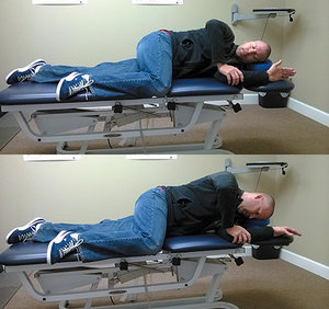 Setup (top) and performance (bottom) positions for the RT2 Supported exercise to increase shoulder IR.
Reinhold recommends having the patient place the scapula flush on the ground, in the scapular plane, to decrease the load and faulty biomechanics placed on the shoulder during the sleeper stretch. However, I suggest eliminating this stretch and using the concepts of "Reflex Turning 2" as described by Vojta.4
Setup (top) and performance (bottom) positions for the RT2 Supported exercise to increase shoulder IR.
Reinhold recommends having the patient place the scapula flush on the ground, in the scapular plane, to decrease the load and faulty biomechanics placed on the shoulder during the sleeper stretch. However, I suggest eliminating this stretch and using the concepts of "Reflex Turning 2" as described by Vojta.4
In a small cohort (n=11) of healthy, young-adult chiropractic students, an average of 11.5 degrees of increased GH internal rotation was observed after performing the "RT2 Supported" exercise. All participants demonstrated an immediate increase in range of motion ranging from 1-21 degrees. Based upon this pilot study, future research is planned to look at both the short-term and long-term effects of this exercise protocol.
An Exercise to Increase GH Internal Rotation
The following details setup and performance of the "RT2 Supported" exercise, as presented by Rich Cohen and based on concepts in movement development seen in the works of Vojta. This exercise activates the abdominal chains. Perform as follows. [See images for setup and performance presentations.]
Setup:
- Have the patient lie on their side, stacked on the shoulder with their down arm at 90 degrees of flexion at the shoulder and elbow. The downside arm will now be in a centrated position.
- The bottom leg should be slightly bent and the top leg placed in the 90-90 position. The top leg should now be centrated. If centration is not obtained, place a support under the bent knee.
- The patient should be lying directly on the shoulder and their head should be supported without any lateral flexion or rotation.
- Place the upside hand on the medial epicondyle and instruct the patient to push down while pronating the downside arm.
- Instruct the patient to attempt to lift themselves off the ground onto their elbow.
- Patient should hold for a 10 second count and repeat 10 times.
Notes:
- Elongation of the spine is key and activation of the lower scapula stabilizers should be felt.
- The neck muscles, levator scapula and trap should be quiet.
- If the exercise seems easy, make sure the top leg is raised to 90 degrees.
- If the exercise seems too difficult, try pushing down harder on the elbow.
Vojta's protocol loads the humeral head, which allows one to roll into a crawling position. This facilitates the raising mechanism of pectoralis, lateral rotators, and abductors of the shoulders. Reflex Turning 2 exercises can help activate the abdominal chains, which will increase the rotation of extremities by restoring the elongation of the spine and the cross-chain patterns of the abdominals.5 Incorporating these principles will likely help restore CNS programming and joint centration, and decrease glenohumeral internal rotation deficits.
References
- Neumann DA. Kinesiology of the Musculoskeletal System: Foundations for Rehabilitation. St. Louis, MO: Mosby Elsevier, 2010.
- Dashottar A. "Posterior Shoulder Tightness Measurements: Differentiating Capsule, Muscle and Bone." Dissertation, Ohio State University, 2012.
- Reinold M. "3 Reasons Why I Don't Use the Sleeper Stretch and Why You Shouldn't Either." MikeReinold.com, July 11, 2011.
- Votja V, Peters A. Votja's Principle. Praha: Grada Publishing, 1995.
- Cohen R. "Introduction to Reflex Locomotion According to Vojta." Lecture, 2010.
For a larger discussion of joint centration, read Dr. Hildebrand's article from the June 17, 2012 issue: "Centration: A Clinical Rehabilitation Approach."
Dr. Justin Hildebrand, a graduate of Cleveland Chiropractic College - Kansas City, practices in Kansas City, Mo., at KC North Spine & Joint Center. Contact him with questions and comments regarding this article via e-mail:
![]() .
.