


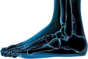
One of the most common conditions of the human frame is excessive foot pronation, in which the foot rolls inward, creating a foot that is flatter, wider and longer. A resultant subluxation pattern of the various tarsals and metatarsals results.
A functional imbalance such as collapse of the arches of the foot also results in excessive motion during the various phases of gait. With excessive pronation, the entire leg spends too much time in internal rotation, placing twisting stresses on the pelvis with each step. Over time, this results in overstretching of the hip and pelvic support ligaments, and can also be a cause of chronic pain and dysfunction.
 An evaluation to check for lower extremity imbalance should be performed on every patient who presents with chronic musculoskeletal conditions of the spine or lower extremities. The following is a recommended series of observations to make while the patient is barefoot and standing:
Foot flare: Toeing out while walking indicates hyperpronation. Weight falls on the medial longitudinal arch, leading to plastic deformation that weakens the foot's supportive qualities.
Knee rotation: Hyperpronation forces the patella to rotate medially, indicating excessive leg movement. The tibia and femur rotate medially, increasing the risk of abnormal hip rotation.
Bowed Achilles tendon(s): The Achilles tendon bows in on the side of hyperpronation. The calcaneus tilts inward, bringing the talus with it. The stress can extend to the tibia and along the entire kinetic chain.
Low medial longitudinal arches: With the patient in a normal, relaxed stance, insert two fingers beneath each medial longitudinal arch. Tight plantar fasciae with possible pain or pressure indicate foot imbalance. As the patient shifts weight outward, note tissue relaxation and absence of pain.
Shoe wear: With hyperpronation, excessive heel wear on the outer edges occurs. Check also for lateral distortion in the counter and/or shoe vamp.2
The 20-30 seconds required to perform the above exam is time well-spent. The information you gather about the feet will be valuable in dealing with postural distortions and pain farther up the body. For example, positive findings often indicate a need for custom-made, flexible foot orthotics to help stabilize the spine and pelvis, and absorb heel-strike shock.
References
- Hyland J, Yochum T, Barry M. "Posture and Weightbearing Biomechanics: Unproved Theory or Clinically Important Concept?" Dynamic Chiropractic, July 29, 1996.
- Charrette M. "Examination of the Foot and Ankle: Non-Weightbearing and Weightbearing Procedures." Success Express, 1996;16(3):20-22.
Click here for previous articles by Mark Charrette, DC.