


Abstract
Spinal deterioration and degeneration are processes that occur in each and every one of our patients. Over time, they lose height because their discs narrow and their curves become twisted or exaggerated.
When the nervous system is dysfunctional or compromised, postural distortions such as upper cervical extension / forward-head posture, increased thoracic kyphosis / barrel chest or pelvic distortion / anterior pelvic tilt occurs.14 These patients can present with a number of pain syndromes such as disc herniations, prolapses, derangements or facet joint arthalgia,12 but no matter their pain, increased spinal compression or load causes degenerative changes.8-9
It is theorized by Schnake, et al., that spinal compression is part of degeneration1 and that care directed at reducing compression through spinal elongation may provide relief for these postural impairments.10-11
According to an article in European Spine Journal,1 "With degeneration, the neutral zone increases in all loading directions while the range of motion decreases for flexion and extension. This may be explained by higher joint laxity. If the potential instability cannot be stabilized by muscles and ligaments sufficiently, loss of spinal balance and increased lordosis are resultant problems."7 Thus, our goal for rehabilitative care is to create decompression through elongation, which can be achieved through traction,10-11 restoring rotation or improving muscular function in the muscles that encourage elongation of the spine.4
An increase in spinal curves in both coronal and sagittal planes shortens the spine and suggests an increase in spinal compression.15 This concept is demonstrated in cases in which scoliotic patients elongate with surgical curve correction.16 As deviations occur, rotation is restricted and compensation, often seen as increased lateral bending, results.
For example, when proximal stability is lost, such as in scapular instability, the distal structures' strength and rotation may be sacrificed.13 According to Passias, et al.,2 when one region is hypomobile, another systematically must compensate and become hypermobile. They showed that when the L5-S1 segment is hypomobile, segments above become hypermobile in flexion and extension. Interestingly, if degeneration is present, the hypermobile segments lose motion in rotation.2
Targeting 3 Muscle Groups to Achieve Elongation
Elongation can be achieved with proper exercises directed at certain muscle groups.4 There are three groups that contribute to the elongation of the spine, and their emphasis in rehabilitation can achieve this goal. The primary spinal elongators are the intrinsic muscles of the spine. Elongation can be accomplished in the cervical spine by activating the deep neck flexors through retraction while uprighting in a cranial direction; and in the lumbar spine by posteriorly tilting the pelvis and drawing the sacrum long.4
The second muscle group that elongates the spine are the abdominals. Activating the transverse abdominalis and oblique abdominal chains in a 360 degree motion will build stability (stiffness) and encourage elongation. As Professor McGill suggests,3 "[T]hose who have more motion in their backs have a greater risk of having future back troubles." One should consider muscle chain exercises to enhance global stability and core activation. Proper core activation increases intra-abdominal pressure and aligns the pelvic floor with the diaphragm.4
A third muscle group to actively elongate the spine is effectively activated by exercising from what Dr. Vojta termed our middle joints: knees and elbows.5 This group consists of the scapular adductors, which include the rhomboids, trapezius and latissimus dorsi. When functioning in a proximal to distal direction, they draw the spine to the distal fixed point. In other words, they become spinal rotators and pull the spinous process toward the distal fix point, effectively elongating the spine.
In this function, the rhomboids, the whole of the trapezius and the lat dorsi act similarly by functioning with a rotational pull on the SPs, so the scapular adductors activate the intrinsic spinal muscles, which then reflexively assist in spinal elongation.6
When exercises are designed to activate the abdominal chains with a punctum fixum at distal fix points, we not only stimulate proper core stabilization, but also activate spinal elongation. For example, when in the side-lying posture, the latissmus dorsi does not work to move the free shoulder, but uses the fixed shoulder or elbow to elongate the spine toward the fixed elbow, and the psoas elongates by rotating the lumbar spinal segments toward the fixed knee.
Additionally, in this position both anterior abdominal chains (cross pattern) are activated, as well as the ipsilateral posterior abdominal chain, further helping to create core stabilization. (See shoulder activation and hip activation exercises below.)
Side-Lying Hip Activation Exercise (Fig. 1)
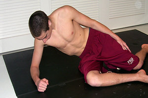 Useful for closed kinetic chain exercises for the hip and pelvis, as well as exercises to elongate the thoracolumbar spine. Also activates the posterior abdominal chain musculature.
Position
- Side-lying on elbow and lateral thigh of lower leg
- Downside elbow with arm to body angle of 90 degrees
- Downside leg positioned with hip, knee and foot at 90 degrees so weight is borne on lateral hip and thigh
- Overside arm is reaching up and forward
- Overside leg is resting on floor with hip and knee at 45 degrees of flexion
Action
- Roll trunk to centrate shoulder and raise off knee as the fulcrum point, lifting the underside hip 2-3 inches off the surface.
- Peel back: no shoulder centration, load support elbow.
- Challenge: raise over knee off floor at 90 degrees of hip flexion, then reduce flexion.
Respiration: Breath normally or in advanced case, use staccato breathing through pursed lips with end-point expiration.
Side-Lying Shoulder Activation Exercise (Fig. 2)
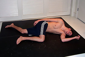 Useful for activation of all shoulder musculature, in particular closed kinetic chain for posterior rotation of the shoulder. Drives glenoid over the humeral head and facilitates muscular function from proximal to distal.
Position: Side-lying on the shoulder with:
- Downside arm trunk angle 90 degrees, elbow less than 90 degrees of flexion, hand open with forearm pronated
- Overside arm reaching to promote an upward and forward movement
- Downside leg resting on surface with hip and knee at 45 degrees of flexion
- Overside leg and knee resting on surface with hip, knee and foot at 90 degrees of flexion
Action
- Raise upper body off the surface approximately 2-3 inches with the downside elbow as the fulcrum point.
- Peel back – support downside elbow with upside hand by pushing down on medial epicondyle.
- Challenge – actively hold upside leg at 90/90 above the surface.
Respiration: Breathe normally or in advanced case, use staccato breathing through pursed lips with end-point expiration.
Evoking spinal elongation creates decompression of the spine by reducing compressive forces on the intervertebral disc, decreasing the load on the spinal facets; and creates rotation in restricted motion segments.10-11 Many commonly treated dysfunctions contribute to degeneration and faulty postural patterns.8-9 It is the design of these exercises to facilitate elongation through muscular traction and further improve stabilization.
References
- Schnake KJ, Putzier M, Haas NP, Kandziora F. Mechanical concepts for disc regeneration. European Spine J, 2006;15(Suppl 3):354-360.
- Passias PG, et al. Segmental lumbar rotation in patients with discogenic low back pain during functional weight-bearing activities. J Bone Joint Surg (U.S.), 2011 Jan 5; 93(1): 29-37.
- McGill SM. Low back exercises: evidence for improving exercise regimens. Physical Ther, 1998;78(7):754-765.
- Kobesova A, Kolar P. Developmental kinesiology: three levels of motor control the assessment and treatment of the motor system. J Bodywork Movement Ther, 2014;18(1):23-33.
- Vojta therapy refresher course, May 2009, Siegen, Germany, under the direction of Wolfram Mueller. PowerPoint lecture with notes.
- Vojta V, Peters A. Das Vojta-Prinzip: Muskelspiele in Reflexfortbewegung Und Motorischer Ontogenese. Heidelberg: Springer, 2007.
- Mimura M, et al. Disc degeneration affects the multidirectional flexibility of the lumbar spine. Spine, 1994;19:1371-1380.
- Lotz JC, Chin JR. Intervertebral disc cell death is dependent on the magnitude and duration of spinal loading. Spine, 2000;25:1477-1483.
- Hsieh AH, Lotz JC. Prolonged spinal loading induces matrix metalloproteinase-2 activation in intervertebral discs. Spine, 2003;28:1781-1788.
- Kroeber M, et al. Effects of controlled dynamic disc distraction on degenerated intervertebral discs. An in vivo study on the rabbit lumbar spine model. Spine, 2005;30:181-187.
- Unglaub F, et al. Controlled distraction as a therapeutic option in moderate degeneration of the intervertebral disc - an in vivo study in the rabbit-spine model. Z Orthop, 2006;144:68-73.
- Urban JPG, Roberts S. Degeneration of the intervertebral disc. Arthritis Res Ther, 2003;5:120-130.
- Sisto SA, Dyson-Hudson T. Dynamometry testing in spinal cord injury. J Rehabil Res Dev, 2007;44(1):123-36.
- Kolar P, Kobesova A. Postural - locomotion function in the diagnosis and treatment of movement disorders, Clin Chiro, 2010;13(1):58-6.
- Stokes IAF, et al. Intervertebral disc changes with angulation, compression and reduced mobility simulating altered mechanical environment in scoliosis. Eur Spine J, 2011 Oct;20(10):1735-1744.
- Hosseini P, et al. Magnetically-controlled growing rods for early onset scoliosis: a multicenter study of 23 cases with minimum 2 years follow-up. Spine, 2016 Mar 10; epub ahead of print.
Dr. Justin Hildebrand, a graduate of Cleveland Chiropractic College - Kansas City, practices in Kansas City, Mo., at KC North Spine & Joint Center. Contact him with questions and comments regarding this article via e-mail:
![]() .
.
Dr. Richard Cohen is a 1985 graduate of Western States Chiropractic College. He is also a diplomate in chiropractic orthopedics, a certified chiropractic sports physician, and board eligible in chiropractic rehabilitation. In addition, Dr. Cohen is credentialed as a McKenzie practitioner and certified Kinesio tape specialist, and is the only DC in the world certified as a Vojta therapist by the International Vojta Society.