


Here's the thing about pain: it comes after inflammation, not before. Pain is the last thing you feel, not the first. In fact, invisible inflammation takes years to manifest itself as pain.
As chiropractors, we pay special attention to the nerves of the body, starting from the spine out. However, we don't always follow the nerve path from beginning to end during hands-on assessment. Inflammation anywhere along the nerve path can cause dysfunction and pain. It's critical to aggressively palpate the nerve pathway to discover underlying inflammation that can cause symptoms far removed from the chief complaint. As Ida Rolf said, "Where you think it is, it ain't."
If you have been treating the site of pain without lasting benefit to the patient, it's time to explore downstream from the spine. Let's look closer at the sciatic nerve and its branches, since lower back pain is such an epidemic.
Anatomy 101: Nerve Roots L4-S3
The sciatic nerve emerges from the lumbosacral plexus. It is the major nerve of the lower limb and the largest nerve in the body, measuring approximately 2 cm. It leaves the pelvis and enters the gluteal region via the greater sciatic foramen of the sacrum.
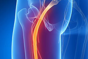 As the nerve moves through the gluteal region, it crosses the posterior surface of the superior gemellus, obturator internus, inferior gemellus and quadratus femoris muscles. It then enters the posterior thigh by passing deep to the long head of the biceps femoris.
Within the posterior thigh, the nerve branches to the hamstring muscles and adductor magnus. When the sciatic nerve reaches the popliteal fossa, it transitions into the tibial and common fibular nerves. The tibial nerve continues its course down the leg, posterior to the tibia. It passes posterior and inferior to the medial malleolus, through the tarsal tunnel.
Assessment Protocols
Now that you understand the anatomy, it's time to assess, starting with visual and then proceeding to manual. The goal in palpation is to be mean when you assess and sweet when you treat. What does that mean? Be forceful in your manual palpation when searching for invisible inflammation and don't cause pain when you treat. Normal tissue should not be painful upon examination. If your patient experiences any pain upon palpation, it is an indication of inflammation. Remember, the inflammation may be far removed from the site of pain.
Visual Assessments: With thepatient prone, notice skin color changes (red, purple, pale, etc). Changes in color indicate superficial nerve changes. Is there a rash or broken blood vessels around an area? This most often indicates an underlying gut issue. Is it puffy looking? The lymphatic system will be involved. Is muscle size different on one side of the lower back, buttock and legs? Atrophy or hypertrophy gives you a glimpse inside the vulnerability and strength of the nervous system.
Palpation: Does the skin feel clammy or dry? Does the skin turn red and stay red when you touch it? These are inflammation markers. Press along and in-between the spinous processes for pain. When assessing the nerve into the lower extremity, it's critical to look at both sides all the way down. Invisible inflammation can settle anywhere in the body and does not have to just be on the symptomatic side.
Forcefully press on the sacral base, apex and sciatic notch. Follow the nerve down under the piriformis into the obturators toward the ischial tuberosity. Severe tenderness may appear when you get close to the obturator internus.
Palpate deep into the belly of a hamstring, noting tissue quality. Can you elicit the jump sign? Notice how deep you can go into the muscle and if the patient experiences muscle guarding on the opposite leg or lower back. Does the patient experience radicular pain with palpation?
One of the most neglected areas of palpation is along the tibial portion of the nerve. Start at the superior midline of the gastrocnemius and follow along the border medial side of the tibia. What can appear as an issue with the soleus muscle or tibialis posterior may actually be inflammation of the tibial nerve.
Feel along the posterior area of the medial malleolus, using direct pressure and cross-friction massage. The withdrawal reflex often occurs when the nerve is inflamed in this region.
What to Do About It
This is where it gets interesting. We already know chiropractic manipulation to the lumbar spine is absolutely critical to restore nerve signalization. Depending on your practice preferences, you may already be using different modalities to treat the lumbosacral spine, such as electric muscle stimulation, ultrasound, laser therapy, etc.
Go back and take note of the areas where you found tenderness and invisible inflammation. Simply add the chosen modality you were doing to the lumbar spine over the areas you found during assessment. For example, let's say you found pain along the posterior part of the medial malleolus. Do non-painful manual therapy stimulation combined with deep-tissue laser therapy to reduce inflammation. Notice the improvement and changes that radiate into the lumbar spine. For an additional powerhouse combination, do the same thing to the opposite-side leg. They all go back to the source.
Top 3 Things to Do After Reading This Article
- Crack open your peripheral nervous system anatomy book or app and familiarize yourself again with the nerve path.
- Start looking at both sides of the body in relationship to nerve irritation. If the right side hurts, also check the left. This applies to all areas of the body.
- Understand there is always invisible inflammation; you just have to find it before pain strikes. The only way to find it is to feel for it.
Resources
- "Diagnostic Anatomy of the Sciatic Nerve." In Russell SM: Examination of Peripheral Nerve Injuries. Thieme Publishers Series, 2006.
- Cook G, et al. Movement: Functional Movement Systems: Screening, Assessment, and Corrective Strategies. Aptos, CA: On Target Publications, 2015.
- Flor H, Turk DC. Pain-related cognitions, pain severity, and pain behaviors in chronic pain patients. Presented at the Fifth World Congress on Pain, International Association for the Study of Pain, Hamburg, 1987.
- Netter FH, Colacino S. Atlas of Human Anatomy. East Hanover, NJ: Navartis, 1997.
Click here for more information about Perry Nickelston, DC, FMS, SFMA.