


Weakness of the intrinsic toe flexors, specifically the flexor digitorum brevis muscle (FDB), is a common and underappreciated cause of a wide range of injuries. In 2015, Sullivan, et al.,1 examined 202 people with chronic heel pain, compared them to 70 asymptomatic people, and determined that weakness of the toe flexors was a common finding in the heel pain group.
Importance of FDB Strength
Because the FDB supports the arch and allows for the transfer of force from the rearfoot to the forefoot during pushoff, maintaining strength in this muscle is important not just for injury prevention, but also for improved athletic performance.
In 2017, Paquette, et al.,3 proved that arch weakness strongly correlates with impaired running performance as we age. More recently, Yuasa, et al.,4 had college athletes perform agility tasks and determined that the quickest athletes had the strongest toes. Because soccer players change direction 700 times in a single game,5 even a slight improvement in toe strength could result in substantial improvements in agility and speed.
Testing FDB Strength
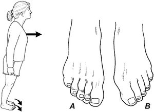 FIG 1 Instruct the standing patient to push their toes into the ground with as much force as possible. A positive wink sign is present when you see a clear crease form at the distal interphalangeal joints (A). In this illustration, the left FDB is weak and there is no visible crease present on the dorsal aspects of the distal interphalangeal joints (B).
While toe strength can be measured with a toe dynamometer and/or the paper grip test, the easiest way to evaluate strength in the FDB is by instructing a standing patient to lean forward and push their toes into the floor with as much force as possible. When the FDB is weak, their second through fifth toes remain stationary, while the distal phalanges plantarflex. (Fig 1A)
FIG 1 Instruct the standing patient to push their toes into the ground with as much force as possible. A positive wink sign is present when you see a clear crease form at the distal interphalangeal joints (A). In this illustration, the left FDB is weak and there is no visible crease present on the dorsal aspects of the distal interphalangeal joints (B).
While toe strength can be measured with a toe dynamometer and/or the paper grip test, the easiest way to evaluate strength in the FDB is by instructing a standing patient to lean forward and push their toes into the floor with as much force as possible. When the FDB is weak, their second through fifth toes remain stationary, while the distal phalanges plantarflex. (Fig 1A)
Conversely, when the FDB is strong, the intermediate phalanges plantarflex, creating a visible crease between the intermediate and distal phalanges, which I refer to as a "positive wink sign." (Fig 1B) A negative or absent wink sign typically indicates weakness not just of the FDB, but also of all the intrinsic muscles of the arch.
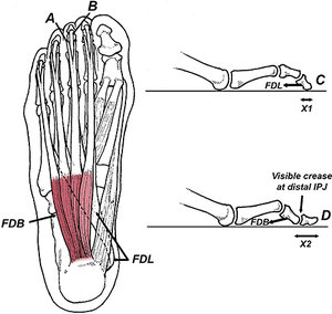 FIG 2 The tendons of FDB bifurcate before attaching to the proximal portion of the intermediate phalanges (A). The flexor digitorum longus muscle (FDL) travels deep to the FDB, and its tendons insert into the bases of the distal phalanges (B). When someone with weakness of the FDB attempts to push their second through fifth toes into the ground, FDL takes over and the distal interphalangeal joints plantarflex (C). In contrast, individuals with strong FDBs create a visible crease along the dorsal aspect of the interphalangeal joints as FDB plantarflexes the proximal interphalangeal joints (D). A strong FDB pulls the distal phalanges directly against the ground, increasing ground contact and the ability to generate force (compare X1 to X2).
The reason the wink sign occurs is because of the interesting relationship between the FDB and flexor digitorum longus (FDL). Just prior to attaching to the intermediate phalanges, the tendons of FDB split, attaching to the plantar medial and lateral sides of the intermediate phalanges. (Fig 2A) The FDL tendons run deep to FDB tendons and attach to the distal phalanges by running directly through the bifurcated FDB tendons. (Fig 2B)
FIG 2 The tendons of FDB bifurcate before attaching to the proximal portion of the intermediate phalanges (A). The flexor digitorum longus muscle (FDL) travels deep to the FDB, and its tendons insert into the bases of the distal phalanges (B). When someone with weakness of the FDB attempts to push their second through fifth toes into the ground, FDL takes over and the distal interphalangeal joints plantarflex (C). In contrast, individuals with strong FDBs create a visible crease along the dorsal aspect of the interphalangeal joints as FDB plantarflexes the proximal interphalangeal joints (D). A strong FDB pulls the distal phalanges directly against the ground, increasing ground contact and the ability to generate force (compare X1 to X2).
The reason the wink sign occurs is because of the interesting relationship between the FDB and flexor digitorum longus (FDL). Just prior to attaching to the intermediate phalanges, the tendons of FDB split, attaching to the plantar medial and lateral sides of the intermediate phalanges. (Fig 2A) The FDL tendons run deep to FDB tendons and attach to the distal phalanges by running directly through the bifurcated FDB tendons. (Fig 2B)
When someone with a weak FDB attempts to plantarflex their toes, the flexor digitorum longus takes over, driving the tips of the toes into the ground. This action results in reduced contact between the ground and the toes, limiting the ability to generate force. (Fig 2C) When the FDB is strong, the intermediate phalanges plantarflex, allowing the toes to make greater ground contact (Fig 2D), which in turn allows for greater force output and enhanced proprioception.
Clinical Implications
The increased digital ground contact present when the FDB is activated may explain why a strong FDB is so important with athletic performance: During terminal pushoff while running or jumping, force is centered beneath the toes, not the metatarsal heads, explaining why the best athletes have the strongest toes.4 In support of this theory, researchers have shown the world's fastest sprinters have toes that are 1 cm longer than recreational runners,6 which provide the FDB and FDL longer lever arms for generating greater force during propulsion.
Keep in mind that while long toes may be necessary for setting world-record sprint times, the implication of this research is that all runners can improve performance simply by strengthening their toes. Toe strength is not just important for sprinting, as Goldman, et al.,7 showed that seven weeks of toe strengthening exercises results in substantial improvements in horizontal jump distance, which is important in a wide range of sports, particularly basketball, volleyball and tennis.
While toe strength is best quantified with a dynamometer, the wink sign represents a quick and easy screen for identifying weakness of the intrinsic muscles of the arch, specifically the FDB. When a positive wink sign is absent, the intrinsic muscles of the arch should be aggressively strengthened.
In addition to improving athletic performance and/or managing plantar fasciitis, toe strengthening exercises have been shown to reduce fall rates in the elderly,8 which is an important cause of disability in people over the age of 70.
References
- Sullivan J, et al. Musculoskeletal and activity-related factors associated with plantar heel pain. Foot & Ankle Int, 2015;36:37-45.
- Abreu M, et al. Plantar calcaneal enthesophytes: new observations regarding sites of origin based on radiographic, MR imaging, anatomic, and paleopathologic analysis. Skeletal Radiol, 2003;32:13-21.
- Paquette M, et al. Biomechanical implications of training volume and intensity in aging runners. Med Sci Sports Exerc, 2018 Mar;50(3):510-515.
- Yuasa Y, et al. Relationship between the toe muscular strength and the ability to change direction in athletes. J Human Kinetics, 2018; 64:47-55.
- Bloomfield J, et al. Physical demands of different positions in FA Premier League soccer. J Sport Sci Med, 2007;6:63-70.
- Lee S, Piazza S. Built for speed: musculoskeletal structure and sprinting ability. J Exper Biol, 2009;212:3700-3707.
- Goldmann J, et al. The potential of toe flexor muscles to enhance performance. J Sport Sci, 2013;31:424-433.
- Mickle K, et al., ISB Clinical Biomechanics Award 2009: toe weakness and deformity increase the risk of falls in older people. Clin Biomech, 2009;24:787-791.
Click here for more information about Thomas Michaud, DC.