


There are several common radiographic findings, seen in the humerus that simulate a pathological process. I'd like to review a few of them as they are relatively common findings.
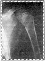
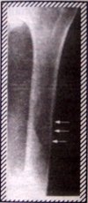
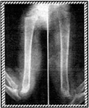
In infants and even adolescents there is often a double contour seen along the lateral or medial metaphyseal region of the humerus, which is a normal finding (Fig. 2). In adults we often see a vertically oriented radiolucent line along the humeral shaft, that is a vascular channel, and may be mistaken for a fracture line. Commonly seen in adults is a radiolucency along the humeral shaft, caused by overlying muscles or soft tissues of the lateral chest wall such as voluminous breats (Fig. 3).
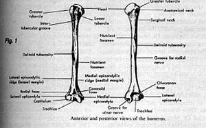
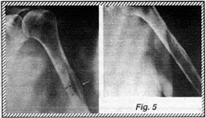
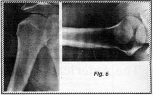
One of the most common questions I am asked is regarding the deltoid tubercle of the humerus. This tubercle is the region of insertion of the deltoid muscle onto the humerus. Generally it is not well visualized, however in some persons it can be very prominent. It generally appears as an irregularity in the contour of the cortex at the lateral proximal aspect of the humeral shaft (Fig. 4). At times however, this area can appear bubbly or cystic, with thickening of the cortex giving the humerus an expanded appearance. Frequently, a strange web-like pattern of sclerotic trabecular densities can be seen just underneath the cortical margin (Fig. 5). Similar changes are found at the insertion of the major pectoralis muscle along the crest of the major tubercle, slightly more proximal to the deltoid tubercle (Fig. 6).
Before alarming the patient, evaluate the humerus with these common radiographic findings in mind. If you're uncertain have a specialist review the study.
Recommended References:
Keats, Atlas of Normal Roentgen Variants.
Kohler/Zimmer, Borderlands of Normal and Early Pathologic Findings in Skeletal Radiography.
Deborah Pate, DC, DACBR
San Diego, California
Click here for more information about Deborah Pate, DC, DACBR.