


Editor's note: The following article expands upon concepts introduced in "Overhead Deep Squats: Understanding Movement and Function," which appeared in the Sept. 28, 2006 issue of DC.
What do you do when your patient with musculoskeletal pain gets 80 percent better and progress seems to be stalled? What's missing to help this patient gain further progress and relief? The answer is in the human movement system. What information are we missing that will allow us to evaluate the human movement system? We can look at how the kinetic chain operates as an integrated functional unit. We need to take a closer look at what our muscles do when we move in everyday life. Functional movements are multidimensional and multiplanar in nature. I find that the deep overhead squat is a useful functional biomechanical analysis.
Current concepts in human movement science provide a useful framework to classify muscle function. We have two distinct yet interdependent muscle systems: the stabilization system (stabilizers or local muscles) and the movement system (mobilizers or global muscles). The local and global muscle systems must integrate for efficient, normal function. Neither system in isolation can control the functional stability of body motion segments. In the presence of chronic or recurrent musculoskeletal pain, patients employ strategies or patterns of muscle recruitment that are normally reserved for high-load function. Pain and pathology do not have to be present concurrently with local muscle dysfunction.
In the presence of pathology and/or pain, a variety of different dysfunctions may present (related to the weak link) in an individual's integrated stability system. Identifying the dysfunction is a priority of treatment. Musculoskeletal pathology can heal and the symptoms may subside; however, the dysfunction does not always automatically return to a normal baseline. The clinical challenge is how to identify the weak link. Commonly accepted methods of identifying the weak link include posture analysis, gait analysis, flexibility assessment, neuromuscular assessment, single-leg balance excursion, multiplanar lunge test, multiplanar step-up test, push-up test, multiplanar vertical jump/hop, multiplanar horizontal jump/hop, shark skill test, multiplanar cone jump/hop test, and speed tests. Other functional tests to assess core stability, strength, and sequencing include the hurdle step and the wall sit with overhead reach. We can divide these assessments for stability and sequencing into static tests, such as drawing-in maneuvers, plank postures, and holding postures in different planes; and dynamic tests, such as Janda's movement patterns. The important thing is to not take out the static tests, but to add dynamic testing to understand the human movement system!
As mentioned in my previous article, the overhead deep squat is a valuable dynamic assessment and exercise. If you wish to exercise the glutes, a full-depth squat is highly recommended. I start my evaluation by saying to the patient, "Just do a squat for me" or "Squat down for me." Observe the patient's natural or normal pattern of movement. Note the feet, knees, hips (lumbar spine), shoulders and the head while the patient performs the squat. After I observe several squats, I ask the patient to squat down while holding the dowel or a barbell over their head. Both elbows should be in the extended position.
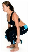
To reiterate, the ideal criteria for a well-performed overhead deep squat are as follows:
- upper torso parallel with the tibia or toward vertical;
- femur below horizontal;
- knees aligned over feet;
- dowel aligned over feet;
- toes pointed forward;
- knees not turned in or out.
Observe: The foot turns outward (externally rotates) while the patient descends. Relate: This implicates a short soleus and gastrocnemius; and long posterior tibialis and medial gastrocnemius. Assess: If the there is excessive outward foot rotation and the hip adducts and/or internally rotates during the descent and/or ascent, this indicates restricted adductors. Rehab solution: Mobilize the external hip rotators; have the patient squat with a spacer between the knees. Place a foam roll or a ball between the patient's knees and have them squeeze the object as they squat. The size of the ball or roll should place the knees approximately shoulder-width apart.
The question often comes up, "Should the knees go over the toes?" The answer is not that it is necessarily wrong, but that it tends to be the way weak people squat. Weak people will exhaust ankle range of motion first and then begin to flex the knee. This results in excessive knee angles or hitting 90 degrees sooner. Think 4:1 knee-to-ankle movement: 4 degrees of knee movement for every 1 degree of ankle movement. If a patient experiences knee pain while doing a squat, they do not necessarily have to be discouraged from performing them. Teach the movement so they are doing the proper technique, loading the correct muscles and joints and not overloading the knees.
The rehab regression for knee pain while squatting is to perform the "airbench" exercise. Have the patient stand against a wall with their feet facing straight ahead; their hips, upper back and head should be against the wall. The patient should walk their feet away from the wall approximately one foot-length; bend their knees and start sliding down the wall until the knees cover the toes as they look down at their feet. Instruct the patient to hold this position and lift the toes upward to keep the weight in their heels; the lower back should be flush up against the wall. Make sure they hold this pose for one to two minutes.
If you have a patient whose chief complaint is low back pain, yet they can do the deep overhead squat and achieve the benchmark of having the upper torso parallel with the tibia or toward the vertical and the femur below horizontal - but the foot flattens, turns outward and the hip abducts - they must stretch the gastrocsoleus complex for improvement within the kinetic chain. This patient can use the overhead deep squat as therapy to correct the tightness in the calf. For rehab, have the patient perform squats with a 1- to 2-inch board under the heels while squatting. Gradually (over many weeks) lower the board until the patient can achieve the benchmark of keeping the feet flat on the ground. Squat repetitions will stretch the tight tissue out.
A method to help stretch tight tissue is postfacilitation technique (PFS) over the gastrocsoleus complex. This is indicated for chronically shortened muscles. The patient performs a maximal contraction with the tight muscle from a midrange position. On relaxation, the doctor quickly stretches the muscle, taking out all the slack. The exact steps for PFS are:
- apply strong force against resistance for approximately seven to 10 seconds;
- instruct the patient to relax immediately;
- elongate the muscle fast and maintain muscle in stretched position (10 to 15 seconds);
- wait approximately 20 seconds before the next resistance, allowing the muscle to regain normal irritability threshold;
- repeat three to five times.
Note: Never stretch if the patient is unable to relax.
A question that often comes up in rehab is, "What should this patient do first, stabilize or mobilize?" Both have significant positive clinical benefits, and it is often advantageous to do both at the same time. The overhead deep squat allows the body to integrate both stability and mobility into function.
If your patient does not meet the benchmark criteria for the overhead deep squat evaluation, you should ask yourself, "Do they need mobilization or stabilization to improve the movement pattern?" The following functional knee-to-chest test will help you sort out this question. Have your patient lie down in the supine position and ask them to "bring the knees to the chest." If they can bring the knees to the chest and maintain a flat back on the floor, they do not have a mobility problem. If you stood them upright on their feet while in the knee-chest position, they should be in the ideal posture for the squat.
Observe: Patient supine - raises arms overhead and performs knee-to-chest. Assess and relate: If the patient has increased ROM, they can do a full squat. It's not a ROM issue. Retest: Challenge the patient for stability versus mobility. Stability is reliably tested under low-load situations. Patient position: Supine; perform bilateral knee-to-chest. Doctor notes the distance and location of the thighs on the torso. Patient's arms are outstretched in front of the body. The practitioner resists bilateral arm flexion. Reassess: Can the patient bring the knees closer to the chest?
Indicates: Increased ROM or decreased pain indicates patient cannot perform the deep overhead squat due to poor stability. Relate: The patient will benefit from a stabilization program. Observe and test: Perform the overhead deep squat with resistance to abduction at the knees (with a band or belt around the knees). Reassess: This leads to increased ROM, but the patient still has pain. Indicates: This is not a gluteus medius issue.
Functional child's pose. Ideally, there should be even flexion throughout the lumbar and thoracic regions. Observe and test: The patient performs a yoga-type child's pose with outstretched arms over the head. Instruct the patient to perform posterior rocking such that the posterior glutes touch the heels. Visually observe the spinal contours. Assess: If a patient has an area of increased kyphosis and is unable to get the spine in a natural curved posture, it is likely a hypomobile or stiff area. The purpose is to assess patients who may be hypomobile. It is important to identify stiff or restricted segments, because these may be a cause of compensatory hypermobility or "give" at an adjacent joint. The site of the "give" or compensation can be the site of the pathology and pain. The stiff area will need to be mobilized with manual techniques and/or the patient can be instructed in the use of a foam roll for self-mobilization.
Improving the quality of the deep overhead squat: Here are four specific progressions and sequences that will improve the deep overhead squat:
Toe-Touch Progression #1:
- Stand erect with feet side by side, heels and toes touching.
- Elevate balls of the feet onto a 1- to 2- inch platform.
- Insert a towel roll or foam roll between the knees.
- Reach for the ceiling, stretching the arms as high as possible with palms facing forward.
- Touch fingertips to toes.
- Repeat for 10 to 12 reps.
Toe-Touch Progression #2:
- Stand erect with feet side by side, heels and toes touching.
- Elevate the heels on a 1- to 2-inch platform; toes on ground.
- Insert a towel roll or foam roll between the knees.
- Reach for the ceiling, stretching the arms as high as possible with palms facing forward.
- Touch fingertips to toes.
- Repeat for 10 to 12 reps.
Reverse Squat Sequence:
- Stand with the heels on a 1- to 2-inch platform, feet spread shoulder-width apart or wider.
- Bend forward until the entire palm can be laid flat on the floor or on a 2-, 4- or 6-inch platform. The entire palm must be flat.
- Lower the body, knees outside of elbows; keep the feet straight.
- Sit deeply into the squat.
- Stretch for 20 seconds.
Deep Squat to a "Y" Position Sequence:
- Start from a deep squat position with the hands on the platform.
- Raise the right arm over the head. Follow the hand with the eyes.
- Raise the left arm over the head. Follow the hand with the eyes.
- With both hands in a "Y" position, extend the spine as much as possible.
- Stand up.
- Repeat for 10 to 12 reps.
How to identify impairment in the overhead deep squat. Observation: The patient complains of flexion-related symptoms in the lumbar spine. The lumbar spine flexes during the descent. Relate: The lumbar spine has greater motion into flexion relative to the hips under flexion load. Rehab: The patient needs to learn to forward lean with a straight back and independent hip flexion, but only as far as the neutral lumbopelvic position can be maintained. Observation: Abnormal lumbar extension during the descent/ascent. Relate: This implicates short illiopsoas, lumbar erectors, quadratus lumborum and latissimus dorsi muscles. Rehab: The patient performs a "single-leg forward bend" with the foot of the tight side on a stool. This puts the knee and hip into slight flexion. Put the foot of the tight side flat on a stationary stool approximately 12 inches high. Ask the patient to bend forward and touch the fingertips to the floor. Repeat this 10-12 times.
 Functional stability grip assist. Observe: During the overhead deep squat, the doctor observes that the heel of foot rises while descending from the neutral starting position. Retest: Ask the patient to keep their feet flat. If you notice a lack of depth with the heels flat on the ground, this may be from a lack of stability and/or a short soleus muscle. Retest: Have the patient perform the "functional stability grip assist deep squat: - the patient grips each hand to a door knob, a bar or the back of a chair. Perform the deep squat. Depth should improve; then stretch the Achilles, gastrocnemius, quadriceps and gluteals. Holding onto a rail or bar will enhance stability that provides active control of the local or global muscle's ability to control motion. Relate: Lack of depth indicates restricted Achilles, gastroc, quads and superficial glut max. Observation: There is a lack of depth and the knees drift internally during descent and/or ascent. Assess: Lack of depth indicates dysfunction of the adductors, gluteals and proximal hamstring. Rehab solution: Lie on your back with your feet up on the wall. As you get more functional, your hips will sit closer to the wall and be flat on the floor at the same time. When you get your legs up on the wall, allow them to spread apart to stretch the adductors. Tighten the thighs and pull your toes back toward your knees and hold for one to four minutes. Your feet must be pointed straight behind you for your hips to be doing the work needed to stabilize the spine. Progression: Perform the above with a foam roll under the lumbar spine to enhance the lordosis. Spread eagle with the legs and feet up along the side of a wall. This will simultaneously stretch the adductors and hamstrings. Observation: The low back goes into flexion during the overhead deep squat. Assess: If the low back goes into flexion to get depth, this implicates the iliotibial band that inserts into the glut max or lack of lumbar control. Solution: Stretch the gluteus and iliotibial region.
Overhead deep squat asymmetry. Observe: Lack of depth or asymmetry occurs during the range of descent motion. Assess: Does the pelvis shift? The pelvis will shift away from restriction. Relate: Asymmetry when squatting indicates restriction in the hip rotators. Rehab: Stretch or mobilize hip rotators. Instruct the patient to lie on their back with both knees bent and their feet flat on the floor, pointed straight ahead. The patient should place their arms out to the side at shoulder level, cross their right ankle over the left knee and rotate the ankle/knee junction in that same direction to the floor. Instruct them to press the right knee away from their body with the right hip musculature; repeat the exercise on the opposite side.
Dysfunction: Asymmetry when squatting shifting to a side. To determine what hip may be causing the dysfunction, check hip height in prone position. Rehab: Stretch and mobilize hips. The prone anterior hip stretch is performed. With the patient in the prone position, place one ankle under the opposite patella. Ideally, the hips should be symmetrical and the height of the anterior hips should be equal distance from the table top. Observe and assess: Asymmetry when squatting shifting to a side indicates weak abductors. Rehab solution: Strengthen the abductors. Perform an abductor exercise by having the patient stand sideways next to the wall. The leg that is closest to the wall should be placed in 90 degrees at the hip and knee. Push with the outside leg into the wall. Progress to wall ball exercises.
Tucker test. As noted in my previous article, the purpose of this test is to help recruit a deeper and stronger contraction of the gluteal group. Test: Place a quarter on the outside of the clothes between the buttocks at the level of the anus and have the patient hold it in place with a strong gluteal contraction. Assess: Can the patient contract the gluteals strong enough and continuously while performing the bridge exercise up and down so the quarter does not drop to the floor? Relate: In order to hold the quarter in place, the patient must concentrate on performing a strong gluteal contraction. This forces the continuous contraction of the gluteus and initiates a co-contraction of the abdominals. Progression: Have the patient perform the overhead deep squat with the quarter held in the buttocks.
Resources
- Bergmark A. Stability of the lumbar spine. A study in mechanical engineering. Acta Orthopaedica Scandinavia 1989;230(60):20-24.
- Caterisano A, Moss RF, Pellinger TK, Woodruff K, Lewis VC, Booth W, Khadra T. The effect of back squat depth on the EMG activity of 4 superficial hip and thigh muscles. J Strength Cond Res August 2002;16(30:428-32
- Cholewicki and McGill. Mechanical stability in the vivo lumbar spine: implications for injury and chronic low back pain. Clinical Biomechanics 1996;11(1):1-15.
- Clark M. "Introduction to Kinetic Chain Dysfunction." Course notes, 2005. Copyright NASM.
- Comerford M. "Lumbo-Pelvic Stability." Course notes, 2003. Copyright M. Comerford.
- Vermeil A. "Sports & Fitness." Course notes, 2005. Copyright A. Vermeil.
- All the coaches, sports medicine specialists and sports scientists who have shared their knowledge with me.
Click here for more information about Jeffrey Tucker, DC, DACRB.